• 文献检索
  • 文档翻译
  • 深度研究
  • 学术资讯
  • Suppr Zotero 插件Zotero 插件
  • 邀请有礼
  • 套餐&价格
  • 历史记录
应用&插件
Suppr Zotero 插件Zotero 插件浏览器插件Mac 客户端Windows 客户端微信小程序
定价
高级版会员购买积分包购买API积分包
服务
文献检索文档翻译深度研究API 文档MCP 服务
关于我们
关于 Suppr公司介绍联系我们用户协议隐私条款
关注我们

Suppr 超能文献

核心技术专利:CN118964589B侵权必究
粤ICP备2023148730 号-1Suppr @ 2026

文献检索

告别复杂PubMed语法,用中文像聊天一样搜索,搜遍4000万医学文献。AI智能推荐,让科研检索更轻松。

立即免费搜索

文件翻译

保留排版,准确专业,支持PDF/Word/PPT等文件格式,支持 12+语言互译。

免费翻译文档

深度研究

AI帮你快速写综述,25分钟生成高质量综述,智能提取关键信息,辅助科研写作。

立即免费体验

使用交错变密度采样和 HighlY 约束笛卡尔重建(HYCR)的肺灌注 MRI。

Pulmonary perfusion MRI using interleaved variable density sampling and HighlY constrained cartesian reconstruction (HYCR).

机构信息

Global Applied Science Laboratory, GE Healthcare, Madison, WI 53705, USA.

出版信息

J Magn Reson Imaging. 2013 Sep;38(3):751-6. doi: 10.1002/jmri.24018. Epub 2013 Jan 24.

DOI:10.1002/jmri.24018
PMID:23349079
原文链接:https://pmc.ncbi.nlm.nih.gov/articles/PMC3638084/
Abstract

PURPOSE

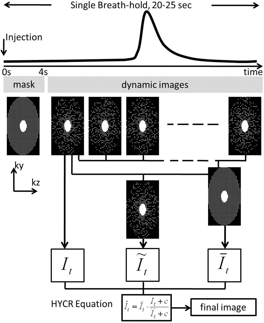
To demonstrate the feasibility of performing single breathhold, noncardiac gated, ultrafast, high spatial-temporal resolution whole chest MR pulmonary perfusion imaging in humans.

MATERIALS AND METHODS

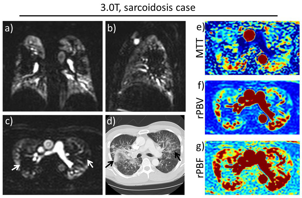
Eight subjects (five male, three female) were scanned with the proposed method on a 3 Tesla clinical scanner using a 32-channel phased-array coil. Seven (88%) were healthy volunteers, and one was a patient volunteer with sarcoidosis. The peak lung enhancement phase for each subject was scored for gravitational effect, peak parenchymal enhancement and severity of artifacts by three cardiothoracic radiologists independently.

RESULTS

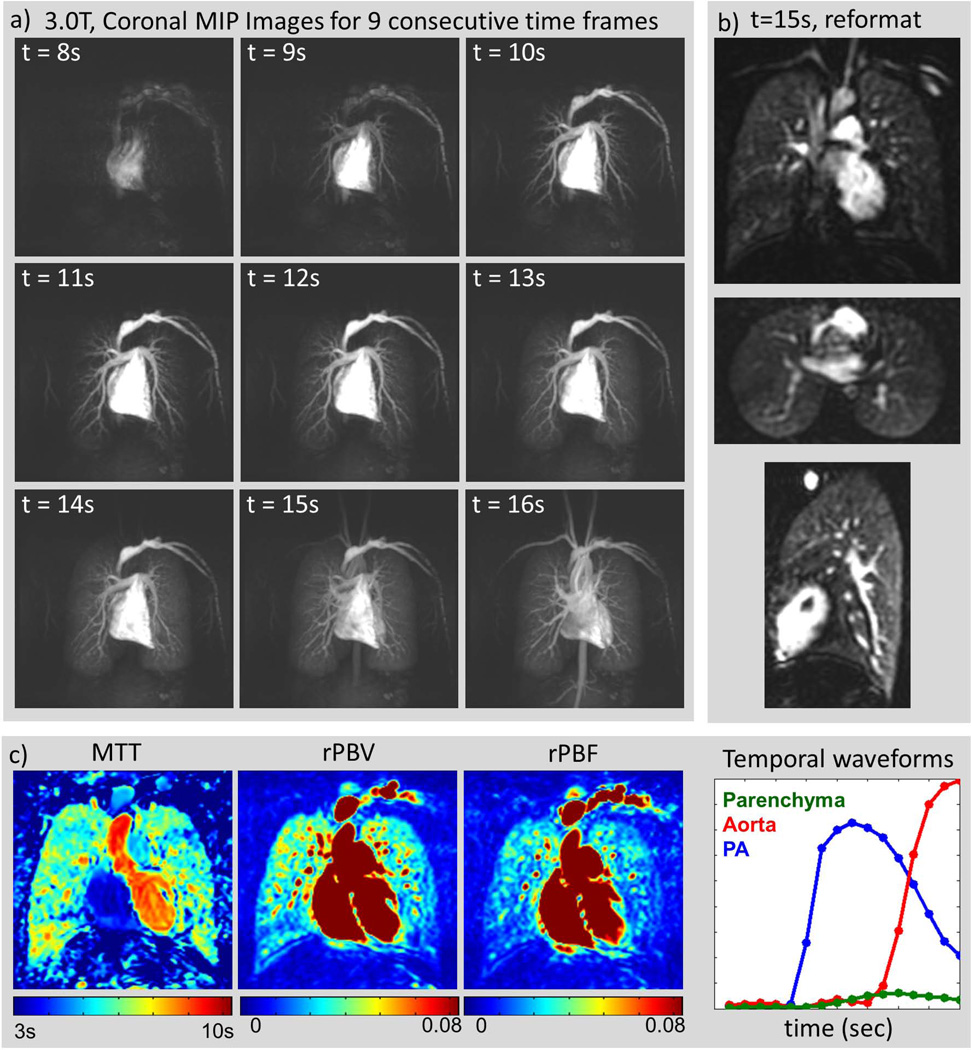
All studies were successfully performed by MR technologists without any additional training. Mean parenchymal signal was very good, measuring 0.78 ± 0.13 (continuous scale, 0 = "none" → 1 = "excellent"). Mean level of motion artifacts was low, measuring 0.13 ± 0.08 (continuous scale, 0 = "none" → 1 = "severe").

CONCLUSION

It is feasible to perform single breathhold, noncardiac gated, ultrafast, high spatial-temporal resolution whole chest MR pulmonary perfusion imaging in humans.

摘要

目的

展示在人体上进行单次屏气、非心脏门控、超快、高时空分辨率全胸部 MR 肺灌注成像的可行性。

材料与方法

8 名受试者(5 名男性,3 名女性)在 3T 临床扫描仪上使用 32 通道相控阵线圈进行了该方法的扫描。其中 7 名(88%)为健康志愿者,1 名患有结节病的患者志愿者。3 名心胸放射科医生独立对每位受试者的肺部增强峰值期的重力效应、实质增强峰值和伪影严重程度进行评分。

结果

所有研究均由磁共振技术人员成功完成,无需额外培训。平均实质信号非常好,测量值为 0.78 ± 0.13(连续量表,0 = “无”→1 = “优秀”)。平均运动伪影水平较低,测量值为 0.13 ± 0.08(连续量表,0 = “无”→1 = “严重”)。

结论

在人体上进行单次屏气、非心脏门控、超快、高时空分辨率全胸部 MR 肺灌注成像的方法是可行的。

https://cdn.ncbi.nlm.nih.gov/pmc/blobs/6b78/3638084/f0e9a92233cb/nihms427273f3.jpg
https://cdn.ncbi.nlm.nih.gov/pmc/blobs/6b78/3638084/30ae167d0530/nihms427273f1.jpg
https://cdn.ncbi.nlm.nih.gov/pmc/blobs/6b78/3638084/b51015ff9c89/nihms427273f2.jpg
https://cdn.ncbi.nlm.nih.gov/pmc/blobs/6b78/3638084/f0e9a92233cb/nihms427273f3.jpg
https://cdn.ncbi.nlm.nih.gov/pmc/blobs/6b78/3638084/30ae167d0530/nihms427273f1.jpg
https://cdn.ncbi.nlm.nih.gov/pmc/blobs/6b78/3638084/b51015ff9c89/nihms427273f2.jpg
https://cdn.ncbi.nlm.nih.gov/pmc/blobs/6b78/3638084/f0e9a92233cb/nihms427273f3.jpg

相似文献

1
Pulmonary perfusion MRI using interleaved variable density sampling and HighlY constrained cartesian reconstruction (HYCR).使用交错变密度采样和 HighlY 约束笛卡尔重建(HYCR)的肺灌注 MRI。
J Magn Reson Imaging. 2013 Sep;38(3):751-6. doi: 10.1002/jmri.24018. Epub 2013 Jan 24.
2
Three-dimensional pulmonary perfusion MRI with radial ultrashort echo time and spatial-temporal constrained reconstruction.采用径向超短回波时间和时空约束重建的三维肺部灌注磁共振成像
Magn Reson Med. 2015 Feb;73(2):555-64. doi: 10.1002/mrm.25158. Epub 2014 Mar 6.
3
All-systolic non-ECG-gated myocardial perfusion MRI: Feasibility of multi-slice continuous first-pass imaging.全收缩期非心电图门控心肌灌注磁共振成像:多层连续首次通过成像的可行性。
Magn Reson Med. 2015 Dec;74(6):1661-74. doi: 10.1002/mrm.25752. Epub 2015 Jun 6.
4
Free-breathing myocardial perfusion MRI using SW-CG-HYPR and motion correction.使用 SW-CG-HYPR 和运动校正的自由呼吸心肌灌注 MRI。
Magn Reson Med. 2010 Oct;64(4):1148-54. doi: 10.1002/mrm.22489.
5
Compressed Sensing for Breast MRI: Resolving the Trade-Off Between Spatial and Temporal Resolution.压缩感知在乳腺 MRI 中的应用:解决空间分辨率和时间分辨率之间的权衡。
Invest Radiol. 2017 Oct;52(10):574-582. doi: 10.1097/RLI.0000000000000384.
6
Improving temporal resolution of pulmonary perfusion imaging in rats using the partially separable functions model.利用部分可分离函数模型提高大鼠肺灌注成像的时间分辨率。
Magn Reson Med. 2010 Oct;64(4):1162-70. doi: 10.1002/mrm.22500.
7
Comprehensive motion-compensated highly accelerated 4D flow MRI with ferumoxytol enhancement for pediatric congenital heart disease.采用菲立磁增强的综合运动补偿高加速4D血流MRI用于小儿先天性心脏病
J Magn Reson Imaging. 2016 Jun;43(6):1355-68. doi: 10.1002/jmri.25106. Epub 2015 Dec 9.
8
Time-resolved 3D pulmonary perfusion MRI: comparison of different k-space acquisition strategies at 1.5 and 3 T.时间分辨三维肺灌注磁共振成像:1.5T和3T下不同k空间采集策略的比较
Invest Radiol. 2009 Sep;44(9):525-31. doi: 10.1097/RLI.0b013e3181b4c252.
9
Highly accelerated cardiac cine phase-contrast MRI using an undersampled radial acquisition and temporally constrained reconstruction.使用欠采样径向采集和时间约束重建的高速心脏电影相位对比 MRI。
J Magn Reson Imaging. 2014 Feb;39(2):455-62. doi: 10.1002/jmri.24160. Epub 2013 Apr 30.
10
Quantification of pulmonary perfusion with free-breathing dynamic contrast-enhanced MRI--a pilot study in healthy volunteers.自由呼吸动态对比增强磁共振成像定量评估肺灌注——一项针对健康志愿者的初步研究
Rofo. 2013 Dec;185(12):1175-81. doi: 10.1055/s-0033-1350128. Epub 2013 Jul 24.

引用本文的文献

1
Dynamic contrast enhanced MRI for the evaluation of lung perfusion in idiopathic pulmonary fibrosis.动态对比增强 MRI 用于特发性肺纤维化肺灌注的评估。
Eur Respir J. 2022 Oct 13;60(4). doi: 10.1183/13993003.02058-2021. Print 2022 Oct.
2
"Structure-Function Imaging of Lung Disease Using Ultrashort Echo Time MRI"."利用超短回波时间 MRI 进行肺部疾病的结构-功能成像"。
Acad Radiol. 2019 Mar;26(3):431-441. doi: 10.1016/j.acra.2018.12.007. Epub 2019 Jan 16.
3
Sparse Reconstruction Techniques in Magnetic Resonance Imaging: Methods, Applications, and Challenges to Clinical Adoption.

本文引用的文献

1
Interleaved variable density sampling with a constrained parallel imaging reconstruction for dynamic contrast-enhanced MR angiography.交错变密度采样结合约束并行成像重建在动态对比增强磁共振血管成像中的应用。
Magn Reson Med. 2011 Aug;66(2):428-36. doi: 10.1002/mrm.22814. Epub 2011 Feb 28.
2
Computed tomography and magnetic resonance imaging of pulmonary hypertension: Pulmonary vessels and right ventricle.肺动脉高压的计算机断层扫描和磁共振成像:肺血管和右心室。
J Magn Reson Imaging. 2010 Dec;32(6):1313-24. doi: 10.1002/jmri.22373.
3
Lung perfusion measured using magnetic resonance imaging: New tools for physiological insights into the pulmonary circulation.
磁共振成像中的稀疏重建技术:方法、应用及临床应用面临的挑战
Invest Radiol. 2016 Jun;51(6):349-64. doi: 10.1097/RLI.0000000000000274.
4
Comparison of models and contrast agents for improved signal and signal linearity in dynamic contrast-enhanced pulmonary magnetic resonance imaging.动态对比增强肺部磁共振成像中用于改善信号及信号线性的模型与对比剂比较
Invest Radiol. 2015 Mar;50(3):174-8. doi: 10.1097/RLI.0000000000000122.
5
Patch based reconstruction of undersampled data (PROUD) for high signal-to-noise ratio and high frame rate contrast enhanced liver imaging.基于补丁的欠采样数据重建(PROUD)用于高信噪比和高帧率对比增强肝脏成像。
Magn Reson Med. 2015 Dec;74(6):1587-97. doi: 10.1002/mrm.25551. Epub 2014 Dec 6.
6
Three-dimensional pulmonary perfusion MRI with radial ultrashort echo time and spatial-temporal constrained reconstruction.采用径向超短回波时间和时空约束重建的三维肺部灌注磁共振成像
Magn Reson Med. 2015 Feb;73(2):555-64. doi: 10.1002/mrm.25158. Epub 2014 Mar 6.
7
Quantitative magnetic resonance imaging of pulmonary hypertension: a practical approach to the current state of the art.肺动脉高压的定量磁共振成像:当前技术水平的实用方法
J Thorac Imaging. 2014 Mar;29(2):68-79. doi: 10.1097/RTI.0000000000000079.
8
Simultaneous MRI of lung structure and perfusion in a single breathhold.在一次屏气过程中对肺结构和灌注进行同步磁共振成像。
J Magn Reson Imaging. 2015 Jan;41(1):52-9. doi: 10.1002/jmri.24520. Epub 2013 Dec 20.
肺灌注磁共振成像测量:探索肺循环生理学的新工具。
J Magn Reson Imaging. 2010 Dec;32(6):1287-301. doi: 10.1002/jmri.22378.
4
Dynamic MR perfusion imaging: capability for quantitative assessment of disease extent and prediction of outcome for patients with acute pulmonary thromboembolism.动态磁共振灌注成像:定量评估急性肺血栓栓塞症患者疾病程度和预测结局的能力。
J Magn Reson Imaging. 2010 May;31(5):1081-90. doi: 10.1002/jmri.22146.
5
Controlled experimental study depicting moving objects in view-shared time-resolved 3D MRA.描述在视图共享时间分辨三维磁共振血管造影中运动物体的对照实验研究。
Magn Reson Med. 2009 Jul;62(1):85-95. doi: 10.1002/mrm.21993.
6
Contrast-enhanced MR angiography using time resolved interleaved projection sampling with three-dimensional Cartesian phase and slice encoding (TRIPPS).使用具有三维笛卡尔相位和切片编码的时间分辨交错投影采样(TRIPPS)的对比增强磁共振血管造影。
Magn Reson Med. 2009 Apr;61(4):918-24. doi: 10.1002/mrm.21805.
7
k-t FOCUSS: a general compressed sensing framework for high resolution dynamic MRI.k-t FOCUSS:一种用于高分辨率动态磁共振成像的通用压缩感知框架。
Magn Reson Med. 2009 Jan;61(1):103-16. doi: 10.1002/mrm.21757.
8
3D high temporal and spatial resolution contrast-enhanced MR angiography of the whole brain.全脑三维高时间和空间分辨率对比增强磁共振血管造影
Magn Reson Med. 2008 Sep;60(3):749-60. doi: 10.1002/mrm.21675.
9
Simulation of relative temporal resolution of time-resolved MRA sequences.时间分辨磁共振血管造影(MRA)序列相对时间分辨率的模拟。
Magn Reson Med. 2008 Aug;60(2):398-404. doi: 10.1002/mrm.21658.
10
3-T MRI for differentiating inflammation- and fibrosis-predominant lesions of usual and nonspecific interstitial pneumonia: comparison study with pathologic correlation.
AJR Am J Roentgenol. 2008 Apr;190(4):878-85. doi: 10.2214/AJR.07.2833.