Laboratory of Molecular Brain Science, Graduate School of Integrated Arts and Sciences, Hiroshima University, Higashi-Hiroshima, Japan.
PLoS One. 2013;8(2):e55559. doi: 10.1371/journal.pone.0055559. Epub 2013 Feb 6.
Estrogen, a class of female sex steroids, is neuroprotective. Estrogen is synthesized in specific areas of the brain. There is a possibility that the de novo synthesized estrogen exerts protective effect in brain, although direct evidence for the neuroprotective function of brain-synthesized estrogen has not been clearly demonstrated. Methylmercury (MeHg) is a neurotoxin that induces neuronal degeneration in the central nervous system. The neurotoxicity of MeHg is region-specific, and the molecular mechanisms for the selective neurotoxicity are not well defined. In this study, the protective effect of de novo synthesized 17β-estradiol on MeHg-induced neurotoxicity in rat hippocampus was examined.
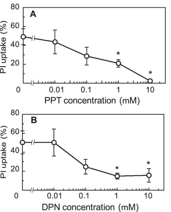
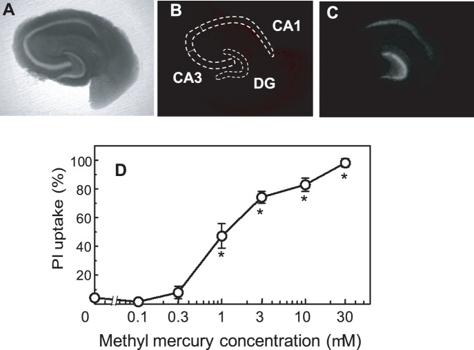
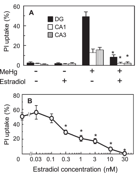
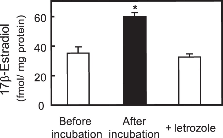
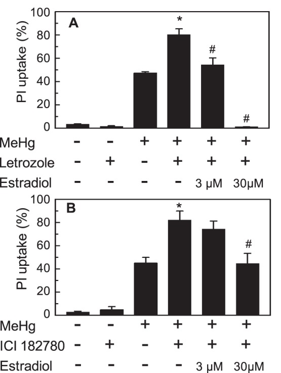
METHODOLOGY/PRINCIPAL FINDINGS: Neurotoxic effect of MeHg on hippocampal organotypic slice culture was quantified by propidium iodide fluorescence imaging. Twenty-four-hour treatment of the slices with MeHg caused cell death in a dose-dependent manner. The toxicity of MeHg was attenuated by pre-treatment with exogenously added estradiol. The slices de novo synthesized estradiol. The estradiol synthesis was not affected by treatment with 1 µM MeHg. The toxicity of MeHg was enhanced by inhibition of de novo estradiol synthesis, and the enhancement of toxicity was recovered by the addition of exogenous estradiol. The neuroprotective effect of estradiol was inhibited by an estrogen receptor (ER) antagonist, and mimicked by pre-treatment of the slices with agonists for ERα and ERβ, indicating the neuroprotective effect was mediated by ERs.
CONCLUSIONS/SIGNIFICANCE: Hippocampus de novo synthesized estradiol protected hippocampal cells from MeHg-induced neurotoxicity via ERα- and ERβ-mediated pathways. The self-protective function of de novo synthesized estradiol might be one of the possible mechanisms for the selective sensitivity of the brain to MeHg toxicity.
雌激素是一类女性性激素,具有神经保护作用。雌激素在大脑的特定区域合成。虽然尚未明确证明脑内合成的雌激素具有神经保护功能,但有可能新合成的雌激素会在大脑中发挥保护作用。甲基汞(MeHg)是一种神经毒素,可在中枢神经系统中诱导神经元变性。MeHg 的神经毒性具有区域特异性,其选择性神经毒性的分子机制尚不清楚。在这项研究中,研究了新合成的 17β-雌二醇对 MeHg 诱导的大鼠海马神经元毒性的保护作用。
方法/主要发现:通过碘化丙啶荧光成像定量检测 MeHg 对海马器官型切片培养物的神经毒性。用 MeHg 处理切片 24 小时可呈剂量依赖性诱导细胞死亡。用外源性添加的雌二醇预处理可减轻 MeHg 的毒性。切片可新合成雌二醇。用 1 μM MeHg 处理不会影响雌二醇的合成。抑制新合成的雌二醇会增强 MeHg 的毒性,而添加外源性雌二醇可恢复毒性增强。雌激素受体(ER)拮抗剂可抑制雌二醇的神经保护作用,并用 ERα和 ERβ激动剂预处理切片可模拟该作用,表明雌二醇的神经保护作用是通过 ER 介导的。
结论/意义:海马新合成的雌二醇通过 ERα 和 ERβ 介导的途径保护海马细胞免受 MeHg 诱导的神经毒性。新合成的雌二醇的自我保护功能可能是大脑对 MeHg 毒性选择性敏感的可能机制之一。