• 文献检索
  • 文档翻译
  • 深度研究
  • 学术资讯
  • Suppr Zotero 插件Zotero 插件
  • 邀请有礼
  • 套餐&价格
  • 历史记录
应用&插件
Suppr Zotero 插件Zotero 插件浏览器插件Mac 客户端Windows 客户端微信小程序
定价
高级版会员购买积分包购买API积分包
服务
文献检索文档翻译深度研究API 文档MCP 服务
关于我们
关于 Suppr公司介绍联系我们用户协议隐私条款
关注我们

Suppr 超能文献

核心技术专利:CN118964589B侵权必究
粤ICP备2023148730 号-1Suppr @ 2026

文献检索

告别复杂PubMed语法,用中文像聊天一样搜索,搜遍4000万医学文献。AI智能推荐,让科研检索更轻松。

立即免费搜索

文件翻译

保留排版,准确专业,支持PDF/Word/PPT等文件格式,支持 12+语言互译。

免费翻译文档

深度研究

AI帮你快速写综述,25分钟生成高质量综述,智能提取关键信息,辅助科研写作。

立即免费体验

抑制 TGF-β 信号通路可促进人眼角膜内皮细胞体外扩增,用于再生医学。

Inhibition of TGF-β signaling enables human corneal endothelial cell expansion in vitro for use in regenerative medicine.

机构信息

Department of Biomedical Engineering, Faculty of Life and Medical Sciences, Doshisha University, Kyotanabe, Japan.

出版信息

PLoS One. 2013;8(2):e58000. doi: 10.1371/journal.pone.0058000. Epub 2013 Feb 25.

DOI:10.1371/journal.pone.0058000
PMID:23451286
原文链接:https://pmc.ncbi.nlm.nih.gov/articles/PMC3581499/
Abstract

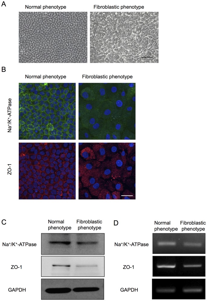
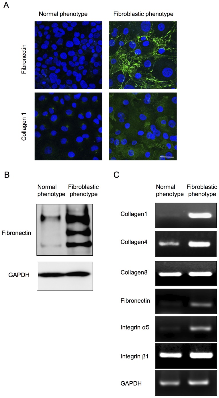
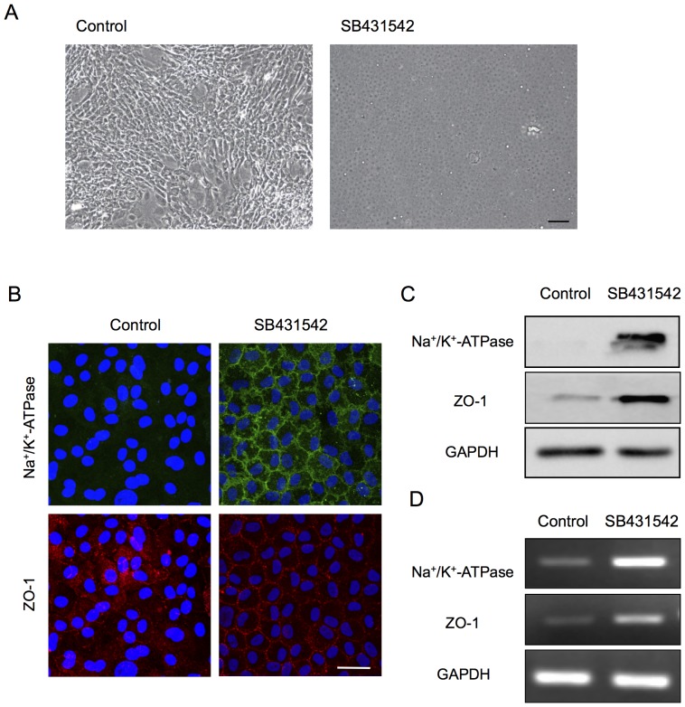
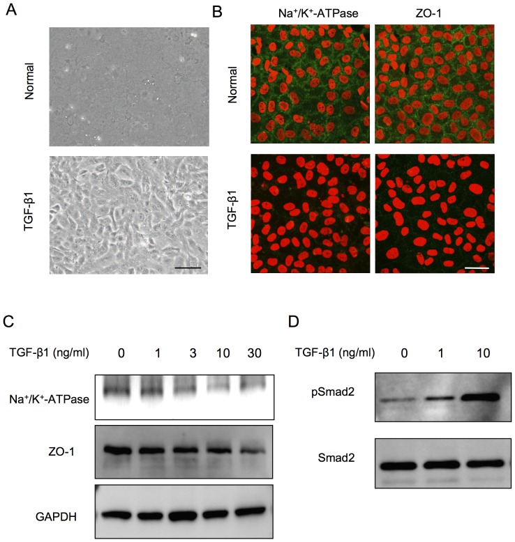
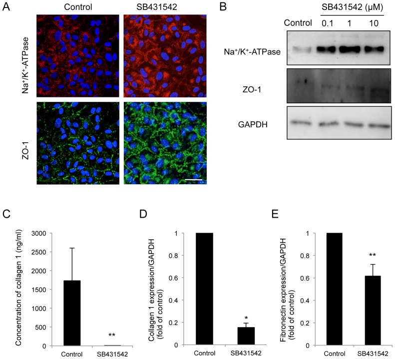
Corneal endothelial dysfunctions occurring in patients with Fuchs' endothelial corneal dystrophy, pseudoexfoliation syndrome, corneal endotheliitis, and surgically induced corneal endothelial damage cause blindness due to the loss of endothelial function that maintains corneal transparency. Transplantation of cultivated corneal endothelial cells (CECs) has been researched to repair endothelial dysfunction in animal models, though the in vitro expansion of human CECs (HCECs) is a pivotal practical issue. In this study we established an optimum condition for the cultivation of HCECs. When exposed to culture conditions, both primate and human CECs showed two distinct phenotypes: contact-inhibited polygonal monolayer and fibroblastic phenotypes. The use of SB431542, a selective inhibitor of the transforming growth factor-beta (TGF-β) receptor, counteracted the fibroblastic phenotypes to the normal contact-inhibited monolayer, and these polygonal cells maintained endothelial physiological functions. Expression of ZO-1 and Na(+)/K(+)-ATPase maintained their subcellular localization at the plasma membrane. Furthermore, expression of type I collagen and fibronectin was greatly reduced. This present study may prove to be the substantial protocol to provide the efficient in vitro expansion of HCECs with an inhibitor to the TGF-β receptor, and may ultimately provide clinicians with a new therapeutic modality in regenerative medicine for the treatment of corneal endothelial dysfunctions.

摘要

在患有 Fuchs 角膜内皮营养不良、假性剥脱综合征、角膜内皮炎和手术引起的角膜内皮损伤的患者中发生的角膜内皮功能障碍会导致失明,因为维持角膜透明性的内皮功能丧失。已经研究了培养角膜内皮细胞 (CEC) 的移植以修复动物模型中的内皮功能障碍,尽管体外扩增人 CEC (HCEC) 是一个关键的实际问题。在这项研究中,我们建立了培养 HCEC 的最佳条件。当暴露于培养条件下时,灵长类动物和人 CEC 都表现出两种截然不同的表型:接触抑制的多边形单层和纤维母细胞样表型。使用 SB431542,一种转化生长因子-β (TGF-β) 受体的选择性抑制剂,可对抗纤维母细胞样表型,使其恢复为正常的接触抑制单层,这些多边形细胞保持了内皮的生理功能。ZO-1 和 Na(+)/K(+)-ATPase 的表达保持其在质膜上的亚细胞定位。此外,I 型胶原和纤维连接蛋白的表达大大减少。本研究可能为提供有效的 TGF-β 受体抑制剂体外扩增 HCEC 提供实质性方案,并最终为临床医生提供一种治疗角膜内皮功能障碍的再生医学新的治疗方法。

https://cdn.ncbi.nlm.nih.gov/pmc/blobs/8e3e/3581499/03ed883bb27a/pone.0058000.g008.jpg
https://cdn.ncbi.nlm.nih.gov/pmc/blobs/8e3e/3581499/edb140340b4f/pone.0058000.g001.jpg
https://cdn.ncbi.nlm.nih.gov/pmc/blobs/8e3e/3581499/0a102383c385/pone.0058000.g002.jpg
https://cdn.ncbi.nlm.nih.gov/pmc/blobs/8e3e/3581499/1190fc175790/pone.0058000.g003.jpg
https://cdn.ncbi.nlm.nih.gov/pmc/blobs/8e3e/3581499/c4034dd7b01f/pone.0058000.g004.jpg
https://cdn.ncbi.nlm.nih.gov/pmc/blobs/8e3e/3581499/f6e349cc2e00/pone.0058000.g005.jpg
https://cdn.ncbi.nlm.nih.gov/pmc/blobs/8e3e/3581499/02e0277bcd25/pone.0058000.g006.jpg
https://cdn.ncbi.nlm.nih.gov/pmc/blobs/8e3e/3581499/46265567df42/pone.0058000.g007.jpg
https://cdn.ncbi.nlm.nih.gov/pmc/blobs/8e3e/3581499/03ed883bb27a/pone.0058000.g008.jpg

相似文献

1
Inhibition of TGF-β signaling enables human corneal endothelial cell expansion in vitro for use in regenerative medicine.抑制 TGF-β 信号通路可促进人眼角膜内皮细胞体外扩增,用于再生医学。
PLoS One. 2013;8(2):e58000. doi: 10.1371/journal.pone.0058000. Epub 2013 Feb 25.
2
LY2109761, Transforming Growth Factor β Receptor Type I and Type II Dual Inhibitor, is a Novel Approach to Suppress Endothelial Mesenchymal Transformation in Human Corneal Endothelial Cells.LY2109761,一种转化生长因子β I型和II型双重抑制剂,是抑制人角膜内皮细胞间充质转化的新方法。
Cell Physiol Biochem. 2018;50(3):963-972. doi: 10.1159/000494480. Epub 2018 Oct 24.
3
Cell surface markers of functional phenotypic corneal endothelial cells.功能性表型角膜内皮细胞的细胞表面标志物。
Invest Ophthalmol Vis Sci. 2014 Nov 11;55(11):7610-8. doi: 10.1167/iovs.14-14980.
4
Involvement of P38MAPK in human corneal endothelial cell migration induced by TGF-β(2).TGF-β(2)诱导的人角膜内皮细胞迁移中 P38MAPK 的作用。
Exp Eye Res. 2013 Mar;108:23-32. doi: 10.1016/j.exer.2012.11.018. Epub 2012 Dec 17.
5
TGF-β1 promotes cell barrier function upon maturation of corneal endothelial cells.TGF-β1 可促进角膜内皮细胞成熟后的细胞屏障功能。
Sci Rep. 2018 Mar 13;8(1):4438. doi: 10.1038/s41598-018-22821-9.
6
Promoting the expansion and function of human corneal endothelial cells with an orbital adipose-derived stem cell-conditioned medium.利用眼眶脂肪来源干细胞条件培养液促进人眼角膜内皮细胞的扩增和功能。
Stem Cell Res Ther. 2017 Dec 20;8(1):287. doi: 10.1186/s13287-017-0737-5.
7
Micro- and nanotopography with extracellular matrix coating modulate human corneal endothelial cell behavior.具有细胞外基质涂层的微米和纳米形貌调节人角膜内皮细胞行为。
Acta Biomater. 2014 May;10(5):1975-84. doi: 10.1016/j.actbio.2014.01.015. Epub 2014 Jan 21.
8
Human Bone Derived Collagen for the Development of an Artificial Corneal Endothelial Graft. In Vivo Results in a Rabbit Model.用于人工角膜内皮移植体开发的人骨源胶原蛋白。兔模型的体内实验结果。
PLoS One. 2016 Dec 1;11(12):e0167578. doi: 10.1371/journal.pone.0167578. eCollection 2016.
9
The Effect of a p38 Mitogen-Activated Protein Kinase Inhibitor on Cellular Senescence of Cultivated Human Corneal Endothelial Cells.p38丝裂原活化蛋白激酶抑制剂对培养的人角膜内皮细胞衰老的影响
Invest Ophthalmol Vis Sci. 2017 Jul 1;58(9):3325-3334. doi: 10.1167/iovs.16-21170.
10
Decellularized bovine corneal posterior lamellae as carrier matrix for cultivated human corneal endothelial cells.去细胞化牛眼角膜后板作为培养的人眼角膜内皮细胞的载体基质。
Curr Eye Res. 2012 Mar;37(3):179-86. doi: 10.3109/02713683.2011.644382.

引用本文的文献

1
SIRT1 Activation Suppresses Corneal Endothelial-Mesenchymal Transition via the TGF-β/Smad2/3 Pathway.SIRT1激活通过TGF-β/Smad2/3信号通路抑制角膜内皮-间充质转化。
Curr Issues Mol Biol. 2024 Dec 6;46(12):13846-13859. doi: 10.3390/cimb46120827.
2
TGF-β-Based Therapies for Treating Ocular Surface Disorders.基于 TGF-β 的眼表疾病治疗方法。
Cells. 2024 Jun 26;13(13):1105. doi: 10.3390/cells13131105.
3
The hiPSC-derived corneal endothelial progenitor-like cell recovers the rabbit model of corneal endothelial dystrophy.

本文引用的文献

1
ROCK inhibitor converts corneal endothelial cells into a phenotype capable of regenerating in vivo endothelial tissue.ROCK 抑制剂将角膜内皮细胞转化为具有体内再生内皮组织能力的表型。
Am J Pathol. 2012 Jul;181(1):268-77. doi: 10.1016/j.ajpath.2012.03.033. Epub 2012 Jun 14.
2
Development of new therapeutic modalities for corneal endothelial disease focused on the proliferation of corneal endothelial cells using animal models.针对角膜内皮细胞增殖的角膜内皮疾病的新治疗方法的开发,使用动物模型。
Exp Eye Res. 2012 Feb;95(1):60-7. doi: 10.1016/j.exer.2011.10.014. Epub 2011 Nov 3.
3
Human corneal endothelial cell expansion for corneal endothelium transplantation: an overview.
人诱导多能干细胞衍生的角膜内皮祖细胞样细胞可恢复角膜内皮营养不良的兔模型。
J Adv Res. 2025 Apr;70:355-369. doi: 10.1016/j.jare.2024.05.008. Epub 2024 May 8.
4
Squishy matters - Corneal mechanobiology in health and disease.黏弹物质——健康与疾病中的角膜生物力学。
Prog Retin Eye Res. 2024 Mar;99:101234. doi: 10.1016/j.preteyeres.2023.101234. Epub 2024 Jan 2.
5
Safety and efficacy of human ESC-derived corneal endothelial cells for corneal endothelial dysfunction.人胚胎干细胞来源的角膜内皮细胞治疗角膜内皮功能障碍的安全性和有效性
Cell Biosci. 2023 Nov 6;13(1):201. doi: 10.1186/s13578-023-01145-w.
6
miR-431 secreted by human vestibular schwannomas increases the mammalian inner ear's vulnerability to noise trauma.人类前庭神经鞘瘤分泌的miR-431增加了哺乳动物内耳对噪声损伤的易感性。
Front Neurol. 2023 Oct 9;14:1268359. doi: 10.3389/fneur.2023.1268359. eCollection 2023.
7
Biomaterials used for tissue engineering of barrier-forming cell monolayers in the eye.用于眼部屏障形成细胞单层组织工程的生物材料。
Front Bioeng Biotechnol. 2023 Sep 27;11:1269385. doi: 10.3389/fbioe.2023.1269385. eCollection 2023.
8
Poly(l-Histidine)-Mediated On-Demand Therapeutic Delivery of Roughened Ceria Nanocages for Treatment of Chemical Eye Injury.聚(组氨酸)介导的粗糙氧化铈纳米笼按需治疗化学性眼损伤。
Adv Sci (Weinh). 2023 Sep;10(26):e2302174. doi: 10.1002/advs.202302174. Epub 2023 Jul 10.
9
A p-Tyr42 RhoA Inhibitor Promotes the Regeneration of Human Corneal Endothelial Cells by Ameliorating Cellular Senescence.一种对酪氨酸42位点的RhoA抑制剂通过改善细胞衰老促进人角膜内皮细胞再生。
Antioxidants (Basel). 2023 May 30;12(6):1186. doi: 10.3390/antiox12061186.
10
Investigating the Role of TGF-β Signaling Pathways in Human Corneal Endothelial Cell Primary Culture.研究 TGF-β 信号通路在人眼角膜内皮细胞原代培养中的作用。
Cells. 2023 Jun 14;12(12):1624. doi: 10.3390/cells12121624.
人眼角膜内皮细胞的扩增用于角膜内皮移植:概述。
Transplantation. 2011 Apr 27;91(8):811-9. doi: 10.1097/TP.0b013e3182111f01.
4
Endothelial keratoplasty - a review.内皮角膜移植术——综述。
Clin Exp Ophthalmol. 2010 Mar;38(2):128-40. doi: 10.1111/j.1442-9071.2010.02213.x.
5
Mechanisms of the epithelial-mesenchymal transition by TGF-beta.TGF-β诱导的上皮-间充质转化的机制。
Future Oncol. 2009 Oct;5(8):1145-68. doi: 10.2217/fon.09.90.
6
Induction of FGF-2 synthesis by IL-1beta in aqueous humor through P13-kinase and p38 in rabbit corneal endothelium.IL-1β 通过 P13-激酶和 p38 在兔角膜内皮细胞中诱导 FGF-2 合成。
Invest Ophthalmol Vis Sci. 2010 Feb;51(2):822-9. doi: 10.1167/iovs.09-4240. Epub 2009 Sep 24.
7
Mitogen activated protein kinases in renal fibrosis.肾纤维化中的丝裂原活化蛋白激酶
Front Biosci (Schol Ed). 2009 Jun 1;1(1):171-87. doi: 10.2741/s17.
8
Enhancement on primate corneal endothelial cell survival in vitro by a ROCK inhibitor.ROCK抑制剂对灵长类动物角膜内皮细胞体外存活的增强作用。
Invest Ophthalmol Vis Sci. 2009 Aug;50(8):3680-7. doi: 10.1167/iovs.08-2634. Epub 2009 Apr 22.
9
Human corneal endothelial cell transplantation in a human ex vivo model.人角膜内皮细胞移植在人离体模型中的应用。
Invest Ophthalmol Vis Sci. 2009 May;50(5):2123-31. doi: 10.1167/iovs.08-2653. Epub 2009 Jan 10.
10
Cultivated corneal endothelial transplantation in a primate: possible future clinical application in corneal endothelial regenerative medicine.灵长类动物中的培养角膜内皮移植:角膜内皮再生医学未来可能的临床应用。
Cornea. 2008 Sep;27 Suppl 1:S48-55. doi: 10.1097/ICO.0b013e31817f2298.