1. Cellular and Molecular Research Center, Tehran University of Medical Science, Tehran, Iran ; 2. Department of Anatomical Sciences, School of Medical Sciences, Tehran University of Medical Sciences, Tehran, Iran.
Cell J. 2012 Summer;14(2):82-9. Epub 2012 Aug 31.
We evaluated structural and functional changes of fresh and frozen-thawed adult mouse spermatogonial stem cells following auto-transplantation into gamma-irradiated testes.
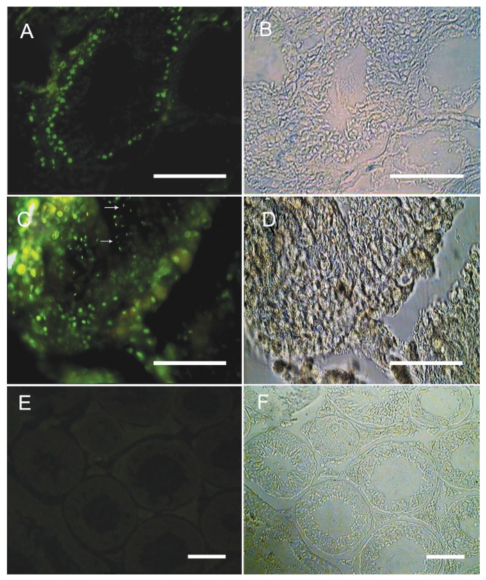
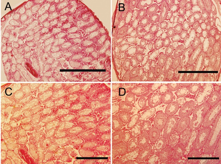
In this experimental research, the right testes from adult mice (n=25) were collected, then Sertoli and spermatogonial cells were isolated using two-step enzymatic digestion, lectin immobilization and differential plating. Three weeks after cultivation, the Bromodeoxyuridine (BrdU)-labeled spermatogonial cells were transplanted, via rete testis, into the other testis of the same mouse, which had been irradiated with 14Gy. The mice were transplanted with: fresh cells (control 1), fresh cells co-cultured with Sertoli cells (control 2), the frozen-thawed cells (experimental 1) and frozen-thawed cells co-cultured with Sertoli cells (experimental 2). The morphological changes between different transplanted testes groups were compared in 8 weeks after transplantation. The statistical significance between mean values was determined by Kruskal Wallis and one-way analysis of variance in efficiency of transplantation.
The statistical analysis revealed significant increases in the mean percentage of testis weight and normal seminiferous tubules following spermatogonial stem cells transplantation in the recipient'fs testes. The normal seminiferous tubules percentage in the co-culture system with fresh cells and frozen-thawed groups were more than those in non-transplanted and fresh cell transplanted groups (p≤0.001).
Our results demonstrated that spermatogonial stem cells in the colonies could result sperm production in the recipient's testes after autologous transplantation.
我们评估了新鲜和冷冻解冻的成年小鼠精原干细胞在自体移植到γ射线照射的睾丸后结构和功能的变化。
在这项实验研究中,收集成年小鼠的右侧睾丸(n=25),然后使用两步酶消化、凝集素固定和差速培养分离支持细胞和精原细胞。培养 3 周后,通过睾丸网将 BrdU 标记的精原细胞移植到同一小鼠的另一个已用 14Gy 照射的睾丸中。将新鲜细胞(对照组 1)、新鲜细胞与支持细胞共培养(对照组 2)、冷冻解冻细胞(实验组 1)和冷冻解冻细胞与支持细胞共培养(实验组 2)分别移植到新鲜细胞和冷冻解冻细胞中。比较移植后 8 周不同移植睾丸组之间的形态变化。采用 Kruskal Wallis 和方差分析比较均值的统计学意义。
统计分析显示,受体睾丸内精原干细胞移植后睾丸重量和正常生精小管的平均百分比显著增加。新鲜细胞和冷冻解冻组共培养系统中的正常生精小管百分比高于未移植和新鲜细胞移植组(p≤0.001)。
我们的结果表明,集落中的精原干细胞在自体移植后可以在受体睾丸中产生精子。