Cellular and Molecular Research Center, Iran University of Medical Sciences, Tehran, Iran ; Department of Anatomical Sciences, School of Medicine, Iran University of Medical Sciences, Tehran, Iran.
Cellular and Molecular Research Center, Iran University of Medical Sciences, Tehran, Iran ; Department of Neurosciences, School of Advanced Medical Technologies, Tehran University of Medical Sciences, Tehran, Iran.
Int J Nanomedicine. 2013;8:4563-76. doi: 10.2147/IJN.S45535. Epub 2013 Nov 27.
A 3D-nanofiber scaffold acts in a similar way to the extracellular matrix (ECM)/basement membrane that enhances the proliferation and self-renewal of stem cells. The goal of the present study was to investigate the effects of a poly L-lactic acid (PLLA) nanofiber scaffold on frozen-thawed neonate mouse spermatogonial stem cells (SSCs) and testis tissues.
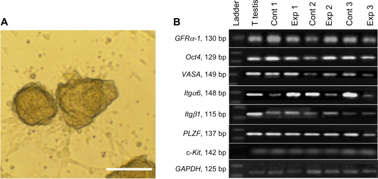
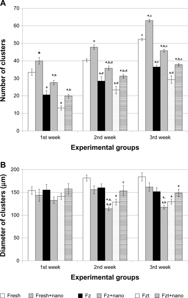
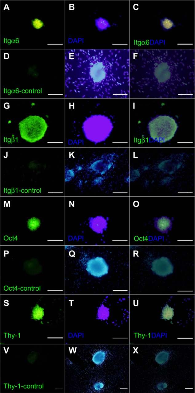
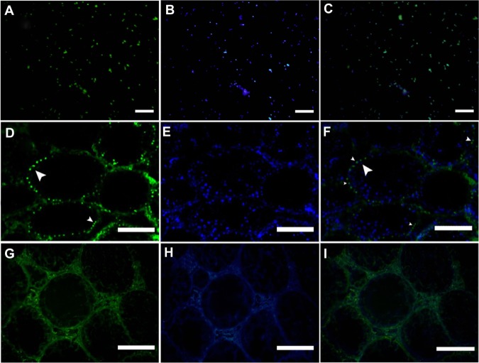
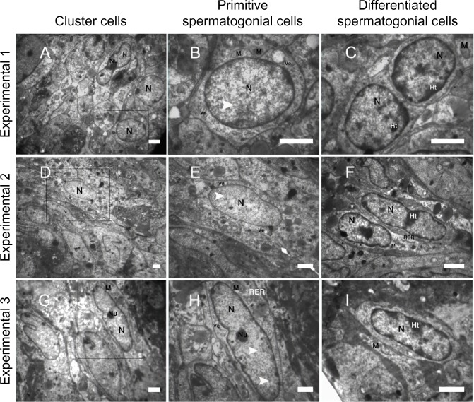
The isolated spermatogonial cells were divided into six culture groups: (1) fresh spermatogonial cells, (2) fresh spermatogonial cells seeded onto PLLA, (3) frozen-thawed spermatogonial cells, (4) frozen-thawed spermatogonial cells seeded onto PLLA, (5) spermatogonial cells obtained from frozen-thawed testis tissue, and (6) spermatogonial cells obtained from frozen-thawed testis tissue seeded onto PLLA. Spermatogonial cells and testis fragments were cryopreserved and cultured for 3 weeks. Cluster assay was performed during the culture. The presence of spermatogonial cells in the culture was determined by a reverse transcriptase polymerase chain reaction for spermatogonial markers (Oct4, GFRα-1, PLZF, Mvh(VASA), Itgα6, and Itgβ1), as well as the ultrastructural study of cell clusters and SSCs transplantation to a recipient azoospermic mouse. The significance of the data was analyzed using the repeated measures and analysis of variance.
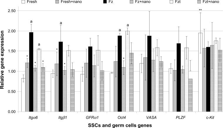
The findings indicated that the spermatogonial cells seeded on PLLA significantly increased in vitro spermatogonial cell cluster formations in comparison with the control groups (culture of SSCs not seeded on PLLA) (P≤0.001). The viability rate for the frozen cells after thawing was 63.00% ± 3.56%. This number decreased significantly (40.00% ± 0.82%) in spermatogonial cells obtained from the frozen-thawed testis tissue. Both groups, however, showed in vitro cluster formation. Although the expression of spermatogonial markers was maintained after 3 weeks of culture, there was a significant downregulation for some spermatogonial genes in the experimental groups compared with those of the control groups. Furthermore, transplantation assay and transmission electron microscopy studies suggested the presence of SSCs among the cultured cells.
Although PLLA can increase the in vitro cluster formation of neonate fresh and frozen-thawed spermatogonial cells, it may also cause them to differentiate during cultivation. The study therefore has implications for SSCs proliferation and germ cell differentiation in vitro.
3D 纳米纤维支架的作用类似于细胞外基质(ECM)/基底膜,可增强干细胞的增殖和自我更新。本研究的目的是研究聚 L-乳酸(PLLA)纳米纤维支架对冷冻解冻新生小鼠精原干细胞(SSC)和睾丸组织的影响。
分离的精原细胞分为六组培养:(1)新鲜精原细胞,(2)接种 PLLA 的新鲜精原细胞,(3)冷冻解冻的精原细胞,(4)接种 PLLA 的冷冻解冻的精原细胞,(5)从冷冻解冻的睾丸组织中获得的精原细胞,(6)从冷冻解冻的睾丸组织中获得的精原细胞接种 PLLA。冷冻保存和培养精原细胞和睾丸组织片段 3 周。在培养过程中进行集落分析。通过逆转录聚合酶链反应检测培养物中精原细胞的存在,检测精原细胞标记物(Oct4、GFRα-1、PLZF、Mvh(VASA)、Itgα6 和 Itgβ1),以及细胞集落的超微结构研究和 SSCs 移植到受体无精子症小鼠中。使用重复测量和方差分析来分析数据的显著性。
研究结果表明,与对照组(未接种 PLLA 的 SSCs 培养)相比,接种 PLLA 的精原细胞在体外精原细胞集落形成中显著增加(P≤0.001)。解冻后细胞的存活率为 63.00%±3.56%。然而,在从冷冻解冻的睾丸组织中获得的精原细胞中,这一数字显著下降(40.00%±0.82%)。两组均能在体外形成集落。尽管培养 3 周后仍保持精原细胞标记物的表达,但与对照组相比,实验组中的一些精原基因表达显著下调。此外,移植实验和透射电子显微镜研究表明,培养细胞中存在 SSCs。
尽管 PLLA 可以增加新生新鲜和冷冻解冻的精原细胞的体外集落形成,但它也可能导致它们在培养过程中分化。因此,该研究对 SSCs 的增殖和体外生殖细胞分化具有重要意义。