Division of Rheumatology, Allergy and Clinical Immunology, University of California at Davis School of Medicine, Davis, CA, USA.
Hepatology. 2013 Sep;58(3):1094-104. doi: 10.1002/hep.26418. Epub 2013 Jul 24.
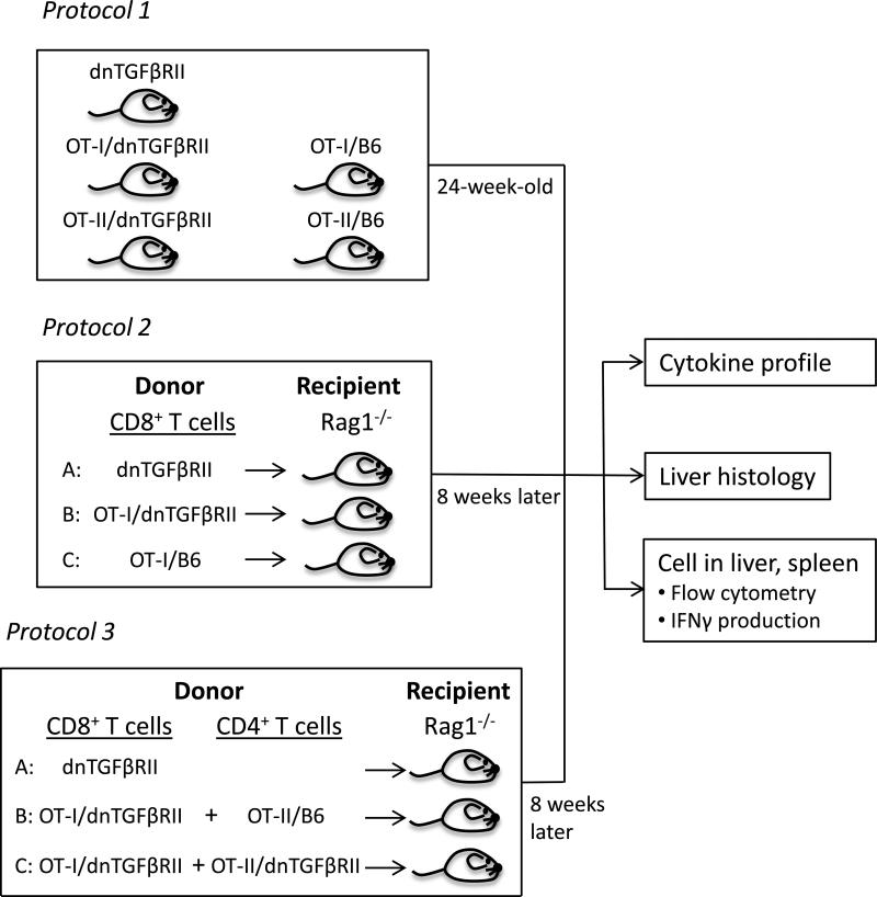
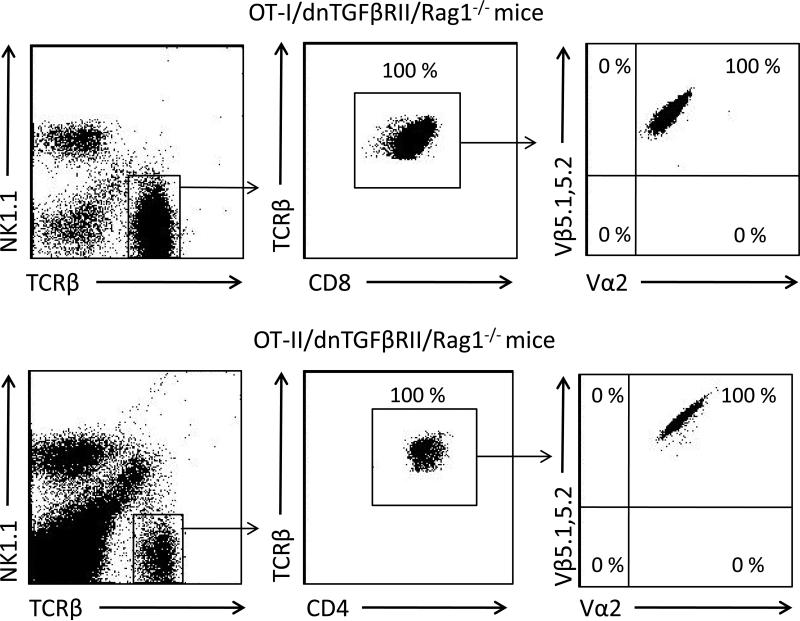
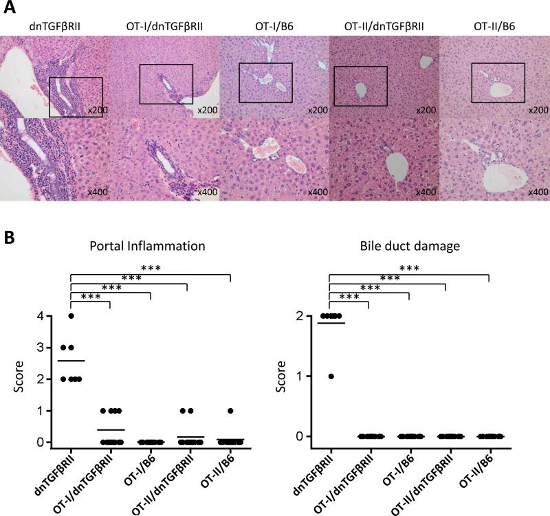
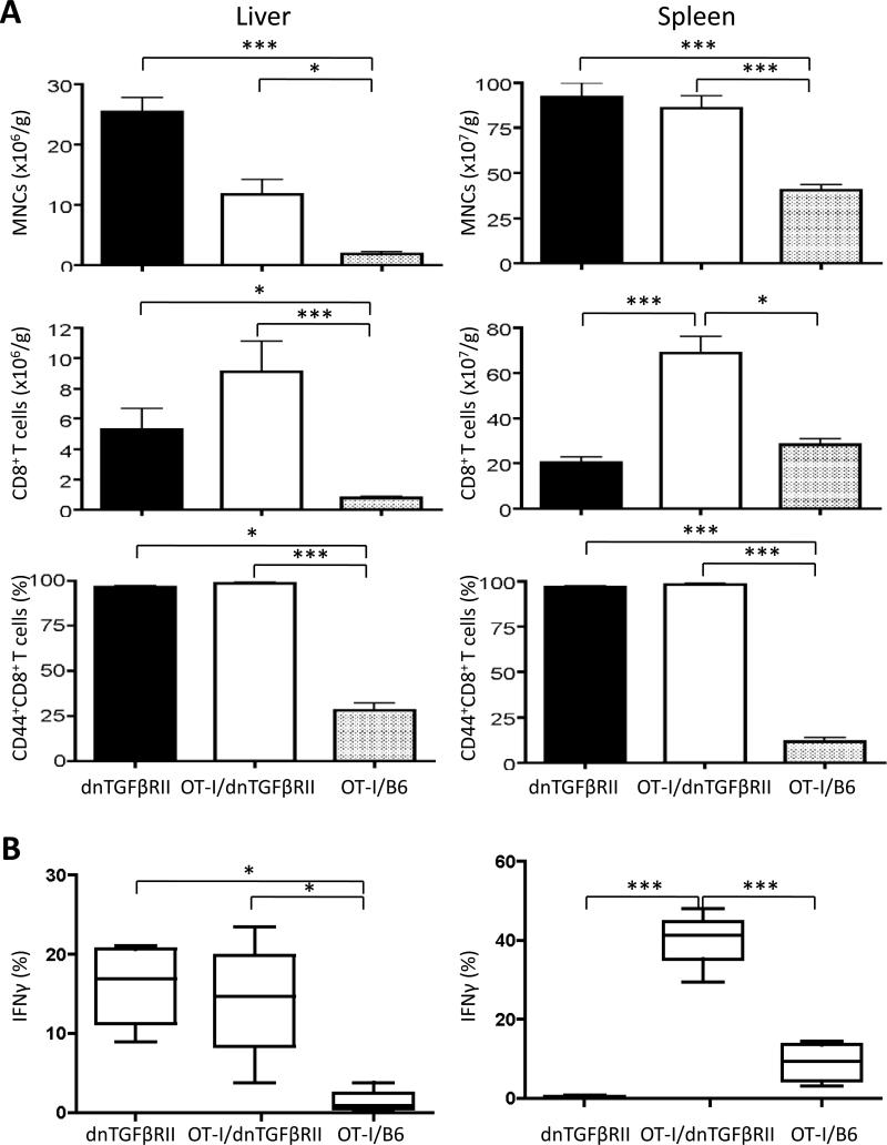
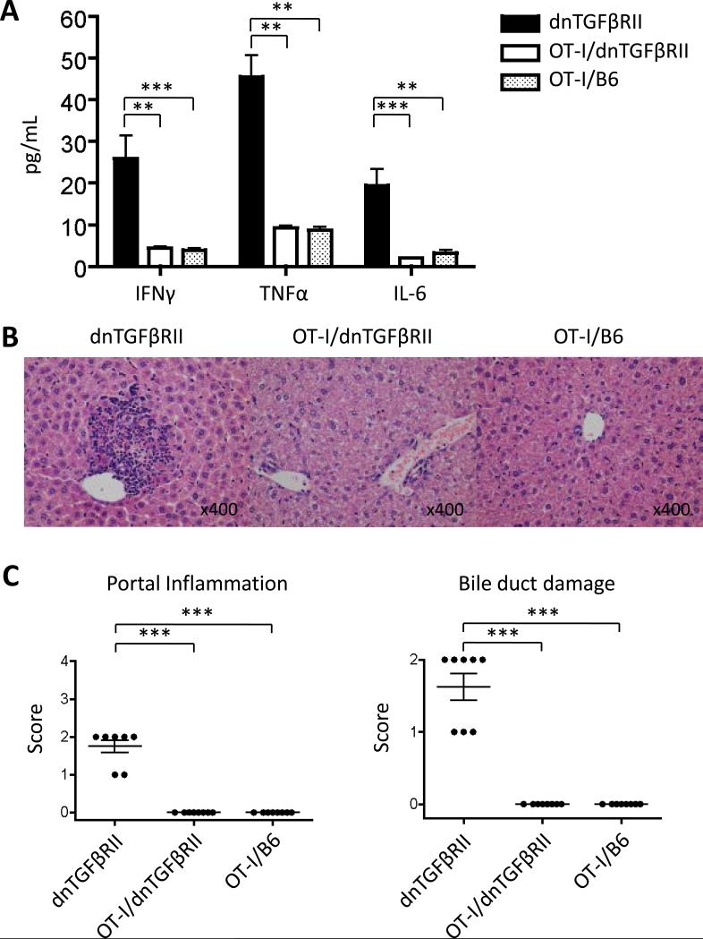
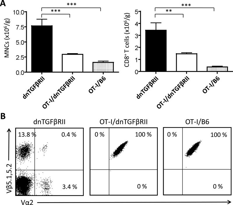
There are several murine models described with features similar to human primary biliary cirrhosis (PBC). Among these models, the one which has the closest serologic features to PBC is a mouse with a T-cell-restricted expression of the dominant negative transforming growth factor β receptor type II (dnTGFβRII). Our work has demonstrated that CD8(+) T cells from dnTGFβRII mice transfer autoimmune cholangitis to Rag1(-/-) recipients. However, it remained unclear whether the autoimmune cholangitis was secondary to an intrinsic function within CD8(+) T cells or due to the abnormal TGFβR environment within which CD8(+) T cells were generated. To address this mechanistic issue, we used our dnTGFβRII, OT-I/Rag1(-/-) , OT-II/Rag1(-/-) mice and in addition generated OT-I/dnTGFβRII/Rag1(-/-) , and OT-II/dnTGFβRII/Rag1(-/-) mice in which the entire T-cell repertoire was replaced with ovalbumin (OVA)-specific CD8(+) or CD4(+) T cells, respectively. Importantly, neither the parental OT-I/dnTGFβRII/Rag1(-/-) mice and/or OT-II/dnTGFβRII/Rag1(-/-) mice developed cholangitis. However, adoptive transfer demonstrated that only transfer of CD8(+) T cells from dnTGFβRII mice but not CD8(+) T cells from OT-I/Rag1(-/-) mice or from OT-I/dnTGFβRII/Rag1(-/-) mice transferred disease. These data were not secondary to an absence of CD4(+) T cell help since a combination of CD8(+) T cells from OT-I/dnTGFβRII/Rag1(-/-) and CD4(+) T cells from OT II/dnTGFβRII/Rag1(-/-) or CD8(+) T cells from OT-I/dnTGFβRII/Rag1(-/-) with CD4(+) T cells from OT-II/Rag1(-/-) mice failed to transfer disease.
Defective TGFβRII signaling, in addition to clonal CD8(+) T cells that target biliary cells, are required for induction of autoimmune cholangitis.
有几种描述的鼠模型具有类似于人类原发性胆汁性肝硬化(PBC)的特征。在这些模型中,与 PBC 的血清学特征最接近的是一种 T 细胞受限表达显性负转化生长因子β受体 II 型(dnTGFβRII)的小鼠。我们的工作表明,dnTGFβRII 小鼠的 CD8+T 细胞可将自身免疫性胆管炎转移给 Rag1(-/-)受体。然而,尚不清楚自身免疫性胆管炎是继发于 CD8+T 细胞的内在功能,还是由于 CD8+T 细胞产生的异常 TGFβR 环境。为了解决这个机制问题,我们使用了 dnTGFβRII、OT-I/Rag1(-/-)、OT-II/Rag1(-/-)小鼠,此外还生成了 OT-I/dnTGFβRII/Rag1(-/-)和 OT-II/dnTGFβRII/Rag1(-/-)小鼠,其中整个 T 细胞库分别被卵清蛋白(OVA)特异性 CD8+或 CD4+T 细胞取代。重要的是,亲本 OT-I/dnTGFβRII/Rag1(-/-)小鼠和/或 OT-II/dnTGFβRII/Rag1(-/-)小鼠均未发生胆管炎。然而,过继转移表明,只有 dnTGFβRII 小鼠的 CD8+T 细胞而非 OT-I/Rag1(-/-)小鼠或 OT-I/dnTGFβRII/Rag1(-/-)小鼠的 CD8+T 细胞可转移疾病。这些数据并不是由于缺乏 CD4+T 细胞辅助所致,因为 OT-I/dnTGFβRII/Rag1(-/-)小鼠的 CD8+T 细胞与 OT-II/dnTGFβRII/Rag1(-/-)或 OT-I/dnTGFβRII/Rag1(-/-)小鼠的 CD4+T 细胞的组合与 OT-II/Rag1(-/-)小鼠的 CD8+T 细胞组合未能转移疾病。
除了针对胆管细胞的克隆性 CD8+T 细胞外,TGFβRII 信号传导缺陷也是诱导自身免疫性胆管炎所必需的。