Li Sha, Bao Hongguang, Han Liu, Liu Lele
Department of Anesthesiology, Nanjing First Hospital Affiliated to Nanjing Medical University, Nanjing 210006, Jiangsu Province, China.
J Biomed Res. 2010 Sep;24(5):389-94. doi: 10.1016/S1674-8301(10)60052-8.
It has been reported that the intravenous anesthetic propofol (PPF) has anti-inflammatory effects in vitro and in patients. The purpose of this study was to investigate whether PPF has anti-inflammatory effects in lipopolysaccharide (LPS)-induced septic shock by inhibiting the induction of pro-inflammatory cytokines [interleukin-6 (IL-6) and tumor necrosis factor-α (TNF-α)] and high mobility group box 1 (HMGB1) in rats.
Thirty six male Wistar rats were randomly assigned to one of three groups (control group, PPF + LPS group and LPS group; n = 12 per group). Control group rats received a 0.9% NaCl solution (NS) by the tail vein. The PPF + LPS group rats received PPF (10 mg/kg bolus, followed by infusion at 10 mg/(kg·h) through a femoral vein catheter) 1 h before LPS (7.5 mg/kg) administration via the tail vein. The LPS group rats received injection of LPS (7.5 mg/kg) via the tail vein. Hemodynamic effects were recorded as well as mortality rates, and plasma cytokine con-centrations (TNF-α, IL-6, HMGB1) were measured for the 24-h observation period.
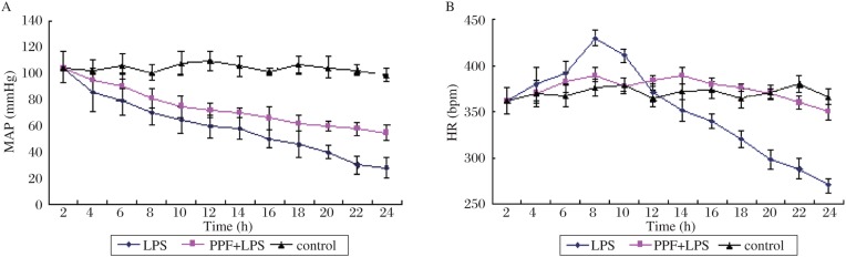
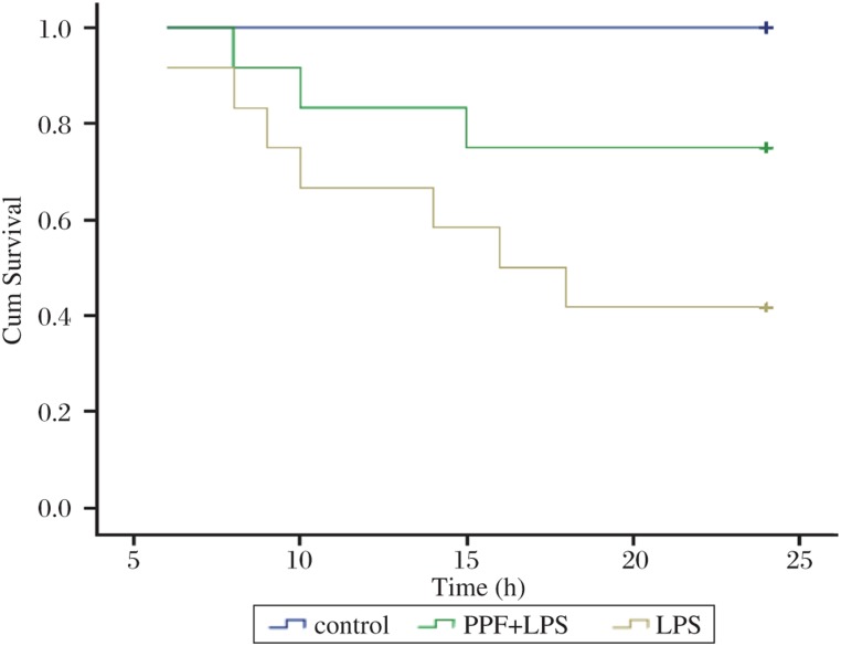
The mean arterial pressure and heart rate of the PPF + LPS group were more stable than those of the LPS group. The mortality at 24 h after the administration of the LPS injection was much higher in the LPS group (58.3%) compared to the PPF + LPS group (25.0%). Plasma concentrations of cytokines (IL-6 and TNF-α) and HMGB1 were significantly reduced in the PPF + LPS group compared to the LPS group (P < 0.05).
Pretreatment with PPF reduced the mortality rate of rats and attenuated the pro-inflammatory cytokine responses in an endotoxin shock model through an anti-inflammatory action inhibiting induction of HMGB1.
据报道,静脉麻醉药丙泊酚(PPF)在体外及患者体内均具有抗炎作用。本研究旨在探讨PPF是否通过抑制大鼠体内促炎细胞因子[白细胞介素-6(IL-6)和肿瘤坏死因子-α(TNF-α)]及高迁移率族蛋白B1(HMGB1)的诱导,从而对脂多糖(LPS)诱导的脓毒症休克产生抗炎作用。
36只雄性Wistar大鼠随机分为三组(对照组、PPF + LPS组和LPS组;每组n = 12)。对照组大鼠经尾静脉给予0.9%氯化钠溶液(NS)。PPF + LPS组大鼠在经尾静脉给予LPS(7.5 mg/kg)前1小时,通过股静脉导管给予PPF(10 mg/kg静脉推注,随后以10 mg/(kg·h)持续输注)。LPS组大鼠经尾静脉注射LPS(7.5 mg/kg)。记录血流动力学效应及死亡率,并在24小时观察期内测定血浆细胞因子浓度(TNF-α、IL-6、HMGB1)。
PPF + LPS组的平均动脉压和心率比LPS组更稳定。LPS注射后24小时,LPS组的死亡率(58.3%)显著高于PPF + LPS组(25.0%)。与LPS组相比,PPF + LPS组的细胞因子(IL-6和TNF-α)及HMGB1的血浆浓度显著降低(P < 0.05)。
在脂多糖休克模型中,PPF预处理通过抑制HMGB1的诱导发挥抗炎作用,降低了大鼠死亡率并减轻了促炎细胞因子反应。