Department of Biology, University of Victoria, British Columbia, V8W 2Y2, Canada.
Nucleic Acids Res. 2013 May;41(9):4888-900. doi: 10.1093/nar/gkt213. Epub 2013 Apr 4.
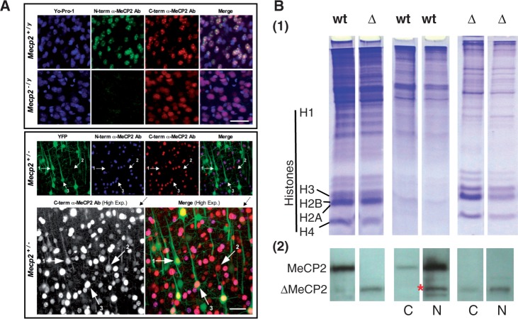
MeCP2 is a methyl-CpG-binding protein that is a main component of brain chromatin in vertebrates. In vitro studies have determined that in addition to its specific methyl-CpG-binding domain (MBD) MeCP2 also has several chromatin association domains. However, the specific interactions of MeCP2 with methylated or non-methylated chromatin regions and the structural characteristics of the resulting DNA associations in vivo remain poorly understood. We analysed the role of the MBD in MeCP2-chromatin associations in vivo using an MeCP2 mutant Rett syndrome mouse model (Mecp2(tm1.1Jae)) in which exon 3 deletion results in an N-terminal truncation of the protein, including most of the MBD. Our results show that in mutant mice, the truncated form of MeCP2 (ΔMeCP2) is expressed in different regions of the brain and liver, albeit at 50% of its wild-type (wt) counterpart. In contrast to the punctate nuclear distribution characteristic of wt MeCP2, ΔMeCP2 exhibits both diffuse nuclear localization and a substantial retention in the cytoplasm, suggesting a dysfunction of nuclear transport. In mutant brain tissue, neuronal nuclei are smaller, and ΔMeCP2 chromatin is digested faster by nucleases, producing a characteristic nuclease-resistant dinucleosome. Although a fraction of ΔMeCP2 is found associated with nucleosomes, its interaction with chromatin is transient and weak. Thus, our results unequivocally demonstrate that in vivo the MBD of MeCP2 together with its adjacent region in the N-terminal domain are critical for the proper interaction of the protein with chromatin, which cannot be replaced by any other of its protein domains.
MECP2 是一种甲基-CpG 结合蛋白,是脊椎动物脑染色质的主要成分。体外研究表明,除了其特定的甲基-CpG 结合结构域(MBD)外,MECP2 还具有几个染色质结合结构域。然而,MECP2 与甲基化或非甲基化染色质区域的特定相互作用以及体内形成的这种 DNA 相互作用的结构特征仍知之甚少。我们使用 Mecp2(tm1.1Jae) (Rett 综合征)突变小鼠模型分析了 MBD 在体内 MECP2-染色质结合中的作用,该模型中 3 号外显子缺失导致蛋白的 N 端截断,包含大多数 MBD。我们的研究结果表明,在突变小鼠中,截短形式的 MECP2(ΔMeCP2)在大脑和肝脏的不同区域表达,尽管只有其野生型(wt)的 50%。与 wt MECP2 的点状核分布特征相反,ΔMeCP2 表现出弥散的核定位和大量保留在细胞质中,这表明核转运功能障碍。在突变脑组织中,神经元核更小,ΔMeCP2 染色质更容易被核酶消化,产生特征性的核酶抗性二核小体。尽管部分 ΔMeCP2 与核小体结合,但与染色质的相互作用是短暂的和弱的。因此,我们的研究结果明确表明,在体内,MECP2 的 MBD 及其 N 端结构域中的相邻区域对于蛋白质与染色质的正确相互作用至关重要,这不能被其任何其他蛋白结构域所替代。