Institute of Biomedical and Biomolecular Science, University of Portsmouth, St Michael's Building, White Swan Rd, Portsmouth, UK.
Neurobiol Dis. 2013 Jul;55(100):87-94. doi: 10.1016/j.nbd.2013.03.016. Epub 2013 Apr 6.
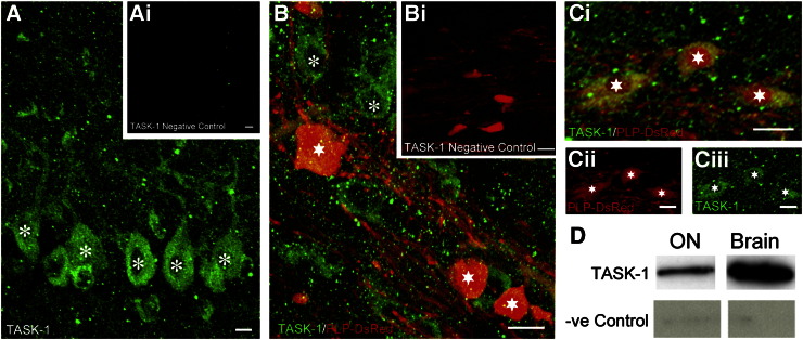
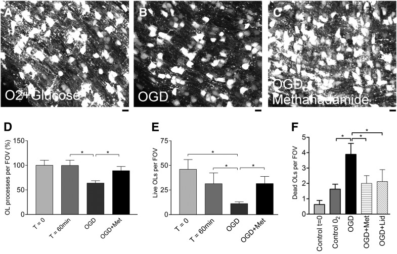
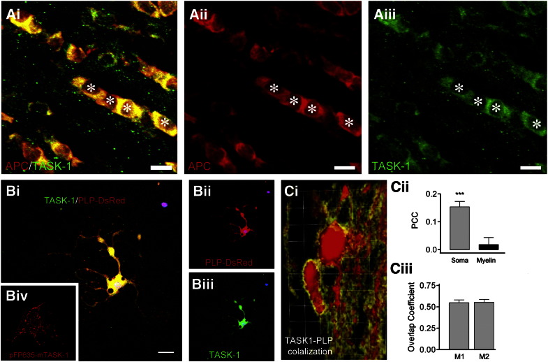
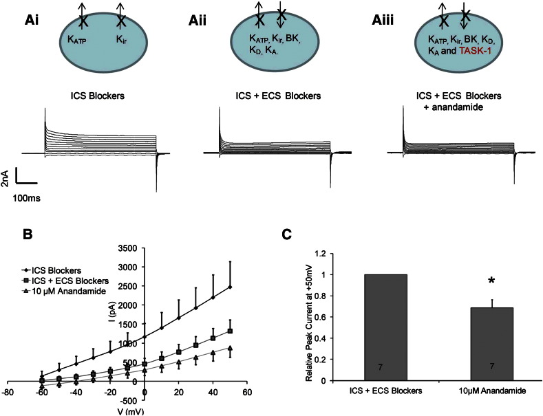
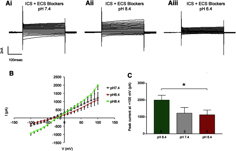
Oligodendrocytes are the myelinating cells of the CNS and, like neurons, are highly sensitive to ischemic damage. However, the mechanisms underlying cytotoxicity in oligodendrocytes during hypoxic/ischemic episodes are not fully understood. TASK-1 is a K(+) leak channel that mediates hypoxic depolarisation in neurons. The expression and function of TASK-1 in oligodendrocytes had not previously been addressed. In this study, we investigate the expression of TASK-1 in oligodendrocytes and its role in white matter ischemic damage. Expression of TASK-1 in oligodendrocytes was investigated in the mouse brain using immunostaining. TASK-1 channel function was identified by established pharmacological and electrophysiological strategies, using the whole-cell patch clamp technique in cell cultures of oligodendrocytes from the optic nerve, a typical white matter tract. The role of TASK-1 in hypoxia was examined in isolated intact optic nerves subjected to oxygen glucose deprivation (OGD). Oligodendrocytes are strongly immunopositive for TASK-1 throughout the brain. Patch-clamp identified functional TASK-1-like leak currents in oligodendrocytes using two recognised means of inhibiting TASK-1, decreasing extracellular pH to 6.4 and exposure to the TASK-1 selective inhibitor anandamide. Incubation of optic nerves with methanandamide, a non-hydrolysable form of anandamide, significantly protected oligodendrocytes against hypoxic disruption and death in OGD. Our data demonstrate for the first time that oligodendrocytes express functional TASK-1 channels and provide compelling evidence they contribute to oligodendrocyte damage in hypoxia. Since oligodendrocyte damage is a key factor in ischemic episodes, TASK-1 may provide a potential therapeutic target in stroke and white matter disease.
少突胶质细胞是中枢神经系统的髓鞘形成细胞,与神经元一样,对缺血性损伤非常敏感。然而,在缺氧/缺血发作期间,少突胶质细胞中细胞毒性的机制尚不完全清楚。TASK-1 是一种介导神经元缺氧去极化的 K(+)渗漏通道。TASK-1 在少突胶质细胞中的表达和功能以前尚未得到解决。在这项研究中,我们研究了 TASK-1 在少突胶质细胞中的表达及其在白质缺血损伤中的作用。使用免疫染色法研究了 TASK-1 在少突胶质细胞中的表达。使用全细胞膜片钳技术在视神经(典型的白质束)的少突胶质细胞培养物中,通过已建立的药理学和电生理策略确定了 TASK-1 通道的功能。使用缺氧葡萄糖剥夺(OGD)法在分离的完整视神经中研究了 TASK-1 在缺氧中的作用。TASK-1 在大脑中广泛表达于少突胶质细胞。使用两种抑制 TASK-1 的方法,即降低细胞外 pH 值至 6.4 和暴露于 TASK-1 选择性抑制剂大麻素,通过膜片钳鉴定了少突胶质细胞中功能性 TASK-1 样渗漏电流。用非水解形式的大麻素甲氧基安非他命孵育视神经可显著保护少突胶质细胞免受 OGD 中的缺氧破坏和死亡。我们的数据首次表明,少突胶质细胞表达功能性 TASK-1 通道,并提供了令人信服的证据表明它们有助于缺氧时少突胶质细胞的损伤。由于少突胶质细胞的损伤是缺血发作的一个关键因素,TASK-1 可能为中风和白质疾病提供一个潜在的治疗靶点。