Onderka D K, Langevin C C, Hanson J A
Alberta Department of Agriculture, Animal Health Division, Edmonton.
Can J Vet Res. 1990 Apr;54(2):285-90.
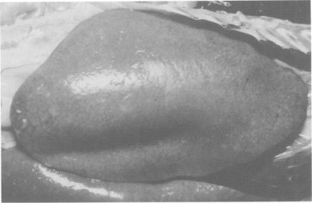
The pathogenesis of fibrosing hepatitis causing condemnations in broiler chickens was investigated. Three to four week old broilers were inoculated via the hepatoenteric bile duct with saline washed suspensions of Clostridium perfringens (10(7) and 10(8) organisms). In another group of broilers, both bile ducts were ligated. The sequential development of liver and gall bladder lesions was studied at intervals ranging from 1-28 days postsurgery. The lesions were similar in both experiments in that the liver became mottled and swollen by five to seven days. Fibrinoid necrosis, heterophil and lymphocyte infiltration, bile duct hyperplasia and fibrosis with reticulin fiber proliferation occurred. By 14-17 days, the liver was enlarged, tan colored and firm with red and white foci. By 28 days, bile duct proliferation and fibrosis were massive with only a few hepatocytes remaining. The liver capsule was not involved. Jaundice was not present but the birds with ligated bile ducts excreted intensely yellow stained droppings after six to seven days. The gall bladder in inoculated birds was edematous and distended with flocculent or inspissated material. Clostridium perfringens was reisolated from gall bladder and/or liver of inoculated birds up to 28 days postsurgery. It is suggested that this organism plays a role in the pathogenesis of fibrosing cholehepatitis by inducing septic intrahepatic cholestasis.
对导致肉鸡被判定不合格的纤维性肝炎的发病机制进行了研究。给3至4周龄的肉鸡通过肝肠胆管接种经盐水洗涤的产气荚膜梭菌悬液(10⁷和10⁸个菌体)。在另一组肉鸡中,双侧胆管被结扎。在手术后1至28天的不同时间间隔研究肝脏和胆囊病变的相继发展情况。两个实验中的病变相似,即肝脏在5至7天时变得斑驳且肿大。出现了纤维蛋白样坏死、嗜异性粒细胞和淋巴细胞浸润、胆管增生以及伴有网状纤维增生的纤维化。到14至17天时,肝脏肿大,呈棕褐色且质地坚实,有红白相间的病灶。到28天时,胆管大量增生和纤维化,仅残留少数肝细胞。肝包膜未受累。未出现黄疸,但胆管结扎的鸡在6至7天后排出深黄色粪便。接种鸡的胆囊水肿且扩张,含有絮状或浓缩物质。在手术后长达28天的时间里,从接种鸡的胆囊和/或肝脏中再次分离出了产气荚膜梭菌。提示该菌通过诱导感染性肝内胆汁淤积在纤维性胆肝炎的发病机制中起作用。