Department of Radiology, Guangzhou First People's Hospital, Guangzhou Medical College, Guangzhou, China.
PLoS One. 2013 Apr 4;8(4):e60279. doi: 10.1371/journal.pone.0060279. Print 2013.
Prospectively assess the performance of diffusion-weighted magnetic resonance imaging (DW-MRI) for differentiation of central lung cancer from atelectasis.
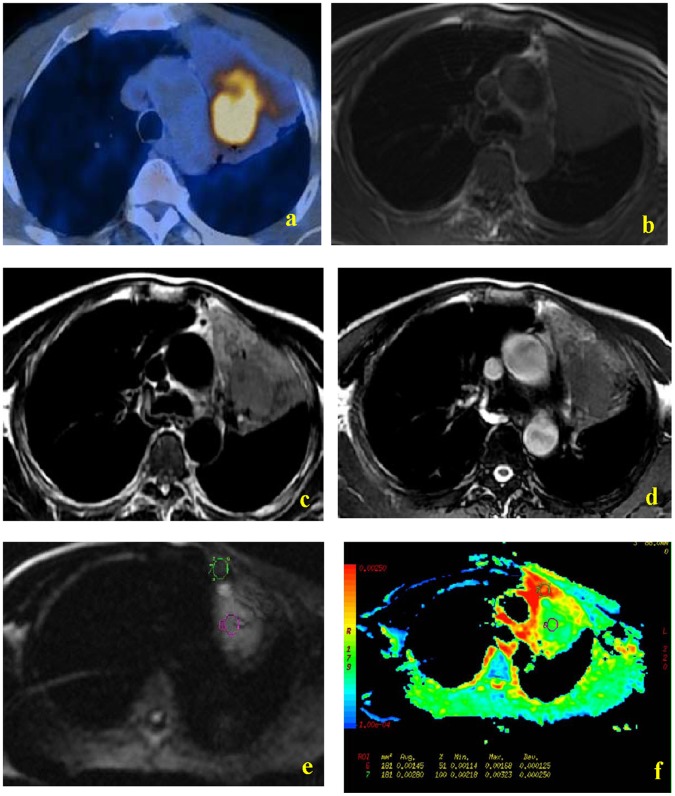
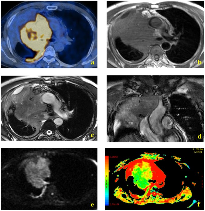
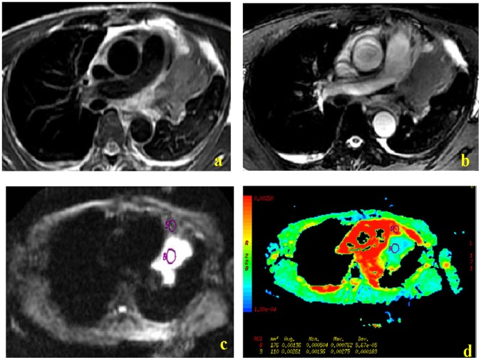
38 consecutive lung cancer patients (26 males, 12 females; age range: 28-71 years; mean age: 49 years) who were referred for thoracic MR imaging examinations were enrolled. MR examinations were performed using a 1.5-T clinical scanner and scanning sequences of T1WI, T2WI, and DWI. Cancers and atelectasis were measured by mapping of the apparent diffusion coefficients (ADCs) obtained with a b-value of 500 s/mm(2).
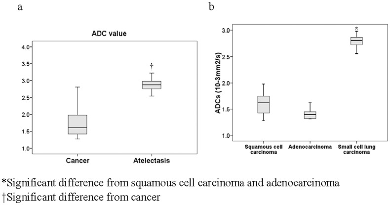
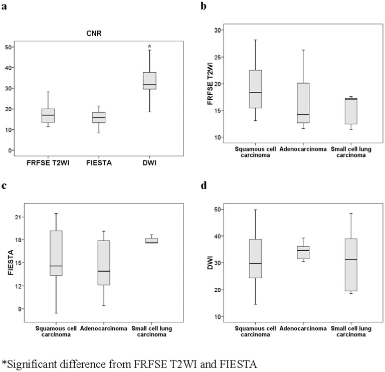
PET/CT and DW-MR allowed differentiation of tumor and atelectasis in all 38 cases, but T2WI did not allow differentiation in 9 cases. Comparison of conventional T2WI and DW-MRI indicated a higher contrast noise ratio of the central lung carcinoma than the atelectasis by DW-MRI. ADC maps indicated significantly lower mean ADC in the central lung carcinoma than in the atelectasis (1.83±0.58 vs. 2.90±0.26 mm(2)/s, p<0.0001). ADC values of small cell lung carcinoma were significantly greater than those from squamous cell carcinoma and adenocarcinoma (p<0.0001 for both).
DW-MR imaging provides valuable information not obtained by conventional MR and may be useful for differentiation of central lung carcinoma from atelectasis. Future developments may allow DW-MR imaging to be used as an alternative to PET-CT in imaging of patients with lung cancer.
前瞻性评估弥散加权磁共振成像(DW-MRI)在中央型肺癌与肺不张鉴别诊断中的性能。
连续纳入 38 例(男 26 例,女 12 例;年龄 28-71 岁,平均年龄 49 岁)因胸部磁共振成像检查而转诊的肺癌患者。使用 1.5-T 临床扫描仪进行磁共振检查,扫描序列包括 T1WI、T2WI 和 DWI。采用 b 值为 500 s/mm2 的表观扩散系数(ADC)图测量癌症和肺不张。
PET/CT 和 DW-MR 可在所有 38 例中区分肿瘤和肺不张,但 T2WI 在 9 例中无法区分。常规 T2WI 和 DW-MRI 的比较表明,DW-MRI 中央型肺癌的对比噪声比高于肺不张。ADC 图显示中央型肺癌的平均 ADC 明显低于肺不张(1.83±0.58 比 2.90±0.26 mm2/s,p<0.0001)。小细胞肺癌的 ADC 值明显大于鳞癌和腺癌(均 p<0.0001)。
DW-MRI 成像提供了常规 MR 无法获得的有价值信息,可能有助于中央型肺癌与肺不张的鉴别诊断。未来的发展可能允许 DW-MRI 成像替代 PET-CT 用于肺癌患者的影像学检查。