Garrison Institute on Aging, Department of Neurology, Texas Tech University Health Sciences Center Lubbock, TX, USA.
Front Aging Neurosci. 2013 May 9;5:19. doi: 10.3389/fnagi.2013.00019. eCollection 2013.
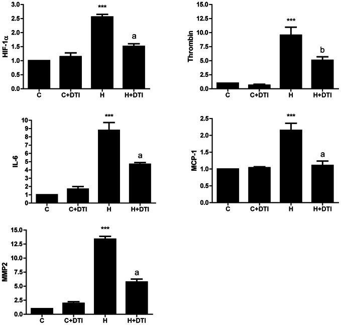
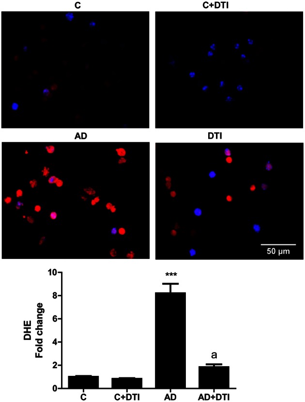
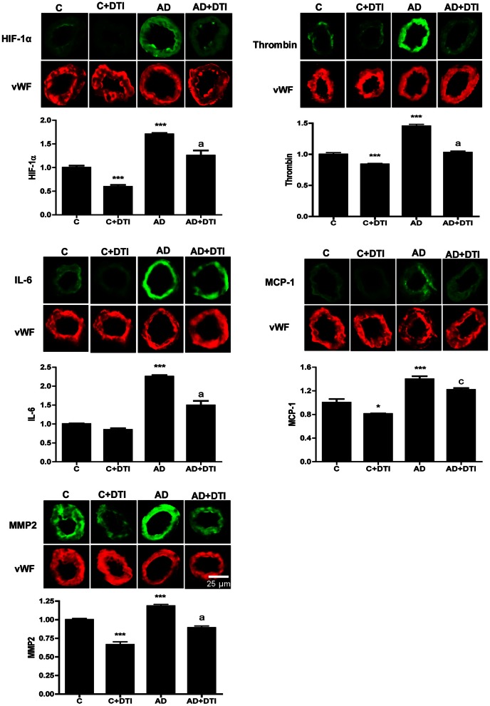
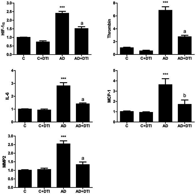
Considerable evidence implicates hypoxia and vascular inflammation in Alzheimer's disease (AD). Thrombin, a multifunctional inflammatory mediator, is demonstrable in the brains of AD patients both in the vessel walls and senile plaques. Hypoxia-inducible factor 1α (HIF-1α), a key regulator of the cellular response to hypoxia, is also upregulated in the vasculature of human AD brains. The objective of this study is to investigate inflammatory protein expression in the cerebrovasculature of transgenic AD mice and to explore the role of thrombin as a mediator of cerebrovascular inflammation and oxidative stress in AD and in hypoxia-induced changes in brain endothelial cells. Immunofluorescent analysis of the cerebrovasculature in AD mice demonstrates significant (p < 0.01-0.001) increases in thrombin, HIF-1α, interleukin-6 (IL-6), monocyte chemoattractant protein-1 (MCP-1), matrix metalloproteinases (MMPs), and reactive oxygen species (ROS) compared to controls. Administration of the thrombin inhibitor dabigatran (100 mg/kg) to AD mice for 34 weeks significantly decreases expression of inflammatory proteins and ROS. Exposure of cultured brain endothelial cells to hypoxia for 6 h causes an upregulation of thrombin, HIF-1α, MCP-1, IL-6, and MMP2 and ROS. Treatment of endothelial cells with the dabigatran (1 nM) reduces ROS generation and inflammatory protein expression (p < 0.01-0.001). The data demonstrate that inhibition of thrombin in culture blocks the increase in inflammatory protein expression and ROS generation evoked by hypoxia. Also, administration of dabigatran to transgenic AD mice diminishes ROS levels in brain and reduces cerebrovascular expression of inflammatory proteins. Taken together, these results suggest that inhibiting thrombin generation could have therapeutic value in AD and other disorders where hypoxia, inflammation, and oxidative stress are involved.
大量证据表明,缺氧和血管炎症与阿尔茨海默病(AD)有关。凝血酶是一种多功能炎症介质,在 AD 患者的大脑中,无论是在血管壁还是在老年斑中,都能被检测到。缺氧诱导因子 1α(HIF-1α)是细胞对缺氧反应的关键调节因子,在人类 AD 大脑的血管中也被上调。本研究旨在研究转基因 AD 小鼠脑血管中的炎症蛋白表达,并探讨凝血酶作为 AD 中血管炎症和氧化应激以及缺氧诱导的脑内皮细胞变化的介质的作用。AD 小鼠脑血管的免疫荧光分析表明,与对照组相比,凝血酶、HIF-1α、白细胞介素-6(IL-6)、单核细胞趋化蛋白-1(MCP-1)、基质金属蛋白酶(MMPs)和活性氧(ROS)的表达显著增加(p < 0.01-0.001)。AD 小鼠连续 34 周给予凝血酶抑制剂达比加群(100mg/kg)治疗,可显著降低炎症蛋白和 ROS 的表达。将培养的脑内皮细胞暴露于缺氧 6 小时会导致凝血酶、HIF-1α、MCP-1、IL-6 和 MMP2 以及 ROS 的上调。内皮细胞用达比加群(1 nM)处理可减少 ROS 的产生和炎症蛋白的表达(p < 0.01-0.001)。这些数据表明,在培养物中抑制凝血酶可阻止缺氧引起的炎症蛋白表达和 ROS 生成的增加。此外,达比加群在 AD 转基因小鼠中的给药可降低大脑中的 ROS 水平并减少脑血管中炎症蛋白的表达。综上所述,这些结果表明,抑制凝血酶的产生可能对 AD 及其他涉及缺氧、炎症和氧化应激的疾病具有治疗价值。