Ozden Raif, Uruc Vedat, Kalacı Aydıner, Dogramacı Yunus
Department of Orthopaedics and Traumatology, Faculty of Medicine, Mustafa Kemal University, Antakya, Hatay, Turkey.
J Brachial Plex Peripher Nerve Inj. 2013 May 30;8(1):5. doi: 10.1186/1749-7221-8-5.
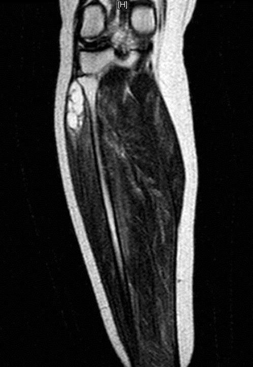
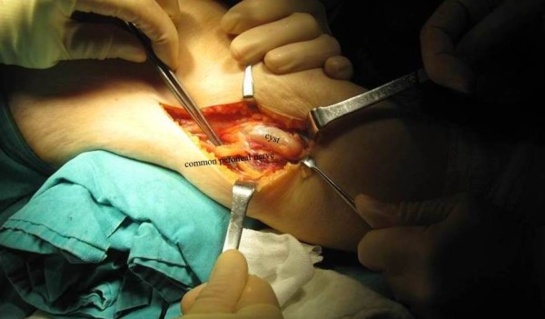
Peripheral neuropathies caused by ganglion cysts are rare. They seldom cause serious complications especially in the lower extremities. The case was a 51-year-old woman referred by her physician to the vascular surgeon with diagnosis including intermittent (vascular) claudication and deep venous thrombosis. Primarily vascular surgeon performed a doppler ultrasound of the lower extremity and calculation of the ankle-brachial index. There were no abnormal pathological findings. Careful physical examination revealed soft swelling and tenderness around the fibular head and neck. Weakness was observed in foot eversion and dorsiflexion. There was pain and tingling in the distribution of the peroneal nerve. and referring the patient to orthopedic surgeon owing to concern for a potential compressive lesion at the right proximal tibiofibular region. Electromyogram studies and physical examination confirmed a diagnosis of compression neuropathy of common peroneal nerve. Magnetic resonance imaging revealed a fluid-filled, lobulated mass indicating a ganglion cyst. One months after decompression, the patient had no complaint. Fast diagnosis and immediate management are essential to regain best possible recovery.
由腱鞘囊肿引起的周围神经病变较为罕见。它们很少引起严重并发症,尤其是在下肢。该病例是一名51岁女性,由其医生转诊至血管外科医生处,诊断包括间歇性(血管性)跛行和深静脉血栓形成。血管外科医生首先对下肢进行了多普勒超声检查并计算了踝肱指数。未发现异常病理结果。仔细的体格检查发现腓骨头和颈部周围有软组织肿胀和压痛。观察到足部外翻和背屈无力。腓总神经分布区域有疼痛和刺痛感,由于担心右近端胫腓区域存在潜在的压迫性病变,将患者转诊至骨科医生处。肌电图检查和体格检查确诊为腓总神经压迫性神经病变。磁共振成像显示一个充满液体的分叶状肿块,提示腱鞘囊肿。减压一个月后,患者无症状。快速诊断和及时处理对于尽可能恢复最佳状态至关重要。