Department of Pathology, Center for Cardiovascular Biology and Institute for Stem Cell and Regenerative Medicine, University of Washington, Seattle, WA 98109, USA.
J Am Heart Assoc. 2013 May 30;2(3):e000202. doi: 10.1161/JAHA.113.000202.
With recent advances in therapeutic applications of stem cells, cell engraftment has become a promising therapy for replacing injured myocardium after infarction. The survival and function of injected cells, however, will depend on the efficient vascularization of the new tissue. Here we describe the arteriogenic remodeling of the coronary vessels that supports vascularization of engrafted tissue postmyocardial infarction (post-MI).
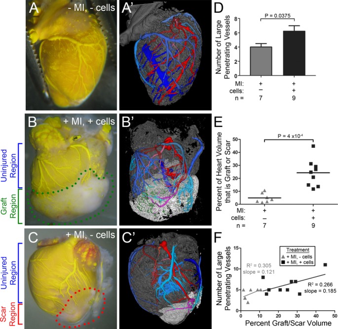
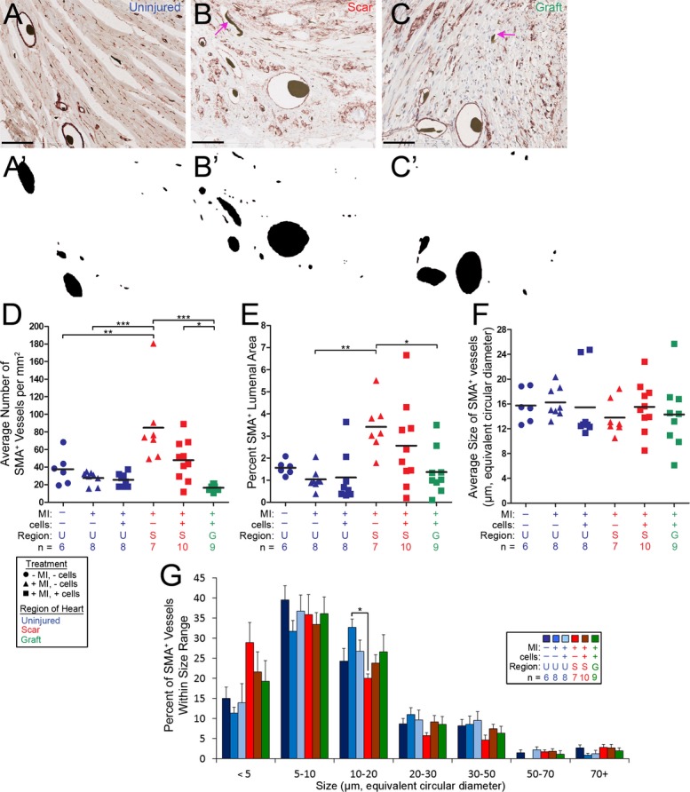
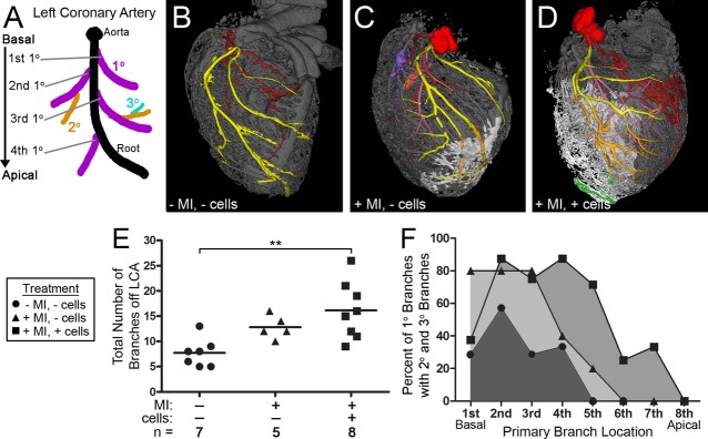
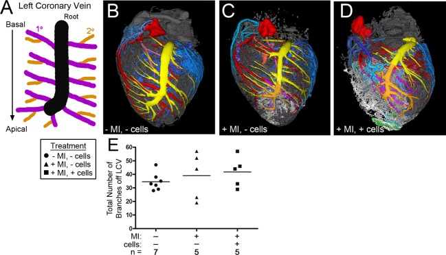
Following MI, murine hearts were injected with a skeletal myoblast cell line previously shown to develop into large grafts. Microcomputed tomography at 28 days postengraftment revealed the 3-dimensional structure of the newly formed conducting vessels. The grafts elicited both an angiogenic response and arteriogenic remodeling of the coronary arteries to perfuse the graft. The coronaries upstream of the graft also remodeled, showing an increase in branching, and a decrease in vascular density. Histological analysis revealed the presence of capillaries as well as larger vascular lumens within the graft. Some graft vessels were encoated by smooth muscle α-actin positive cells, implying that vascular remodeling occurs at both the conducting arterial and microvascular levels.
Following MI and skeletal myoblast engraftment, the murine coronary vessels exhibit plasticity that enables both arteriogenic remodeling of the preexisting small branches of the coronary arteries and development of large and small smooth muscle encoated vessels within the graft. Understanding the molecular mechanisms underlying these 2 processes suggests mechanisms to enhance the therapeutic vascularization in patients with myocardial ischemia.
随着干细胞治疗应用的最新进展,细胞移植已成为一种有前途的治疗方法,可用于替代梗死后受损的心肌。然而,注入细胞的存活和功能将取决于新组织的有效血管化。在这里,我们描述了支持心肌梗死后(梗死后)移植组织血管化的冠状动脉的动脉生成重塑。
梗死后,将先前显示可发育成大移植物的骨骼肌成肌细胞系注射到小鼠心脏中。移植后 28 天的微计算机断层扫描显示了新形成的传导血管的三维结构。移植物引发了血管生成反应和冠状动脉的动脉生成重塑,以灌注移植物。移植物上游的冠状动脉也进行了重塑,表现为分支增加和血管密度降低。组织学分析显示移植物内存在毛细血管和更大的血管腔。一些移植物血管被平滑肌α-肌动蛋白阳性细胞包裹,这意味着血管重塑发生在传导动脉和微血管水平。
梗死后和骨骼肌成肌细胞移植后,小鼠冠状动脉表现出可塑性,既能使冠状动脉的原有小分支发生动脉生成重塑,又能在移植物内形成大和小的平滑肌包裹血管。了解这两个过程背后的分子机制提示了增强心肌缺血患者治疗性血管化的机制。