文献检索文档翻译深度研究
Suppr Zotero 插件Zotero 插件
邀请有礼套餐&价格历史记录

新学期,新优惠

限时优惠:9月1日-9月22日

30天高级会员仅需29元

1天体验卡首发特惠仅需5.99元

了解详情
不再提醒
插件&应用
Suppr Zotero 插件Zotero 插件浏览器插件Mac 客户端Windows 客户端微信小程序
高级版
套餐订阅购买积分包
AI 工具
文献检索文档翻译深度研究
关于我们
关于 Suppr公司介绍联系我们用户协议隐私条款
关注我们

Suppr 超能文献

核心技术专利:CN118964589B侵权必究
粤ICP备2023148730 号-1Suppr @ 2025

不同类型皮肤利什曼病中利什曼原虫皮肤试验反应的评估。

Evaluation of leishmanin skin test reaction in different variants of cutaneous leishmaniasis.

作者信息

Sadeghian Giti, Ziaei Hengameh, Bidabadi Leila Shirani, Nilforoushzadeh Mohammad Ali

机构信息

Department of Skin Diseases and Leishmaniasis Research Center, Isfahan University of Medical Sciences, Isfahan, Iran.

出版信息

Indian J Dermatol. 2013 May;58(3):239. doi: 10.4103/0019-5154.110838.


DOI:10.4103/0019-5154.110838
PMID:23723480
原文链接:https://pmc.ncbi.nlm.nih.gov/articles/PMC3667292/
Abstract

INTRODUCTION: 
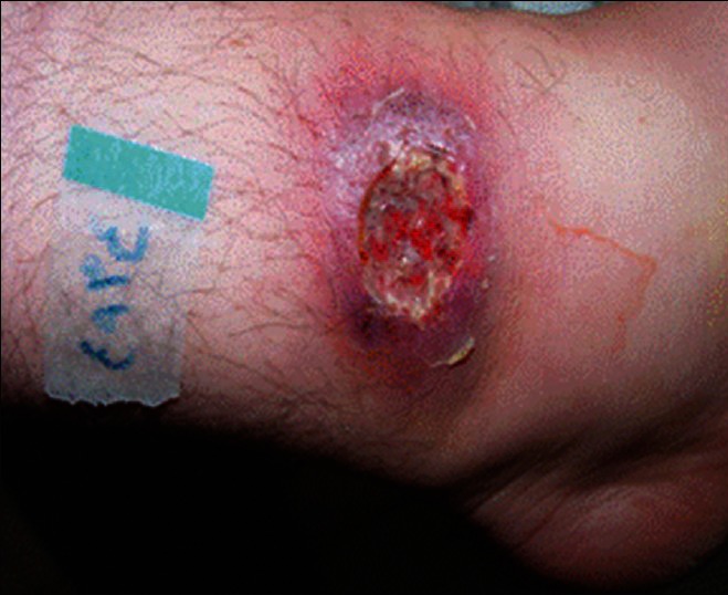
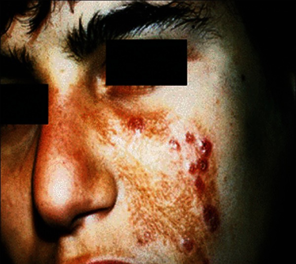
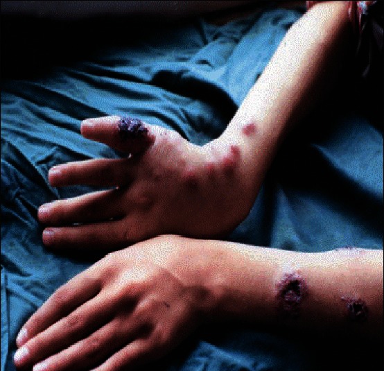
Cutaneous leishmaniasis (CL) is a parasitic disease which has different clinical forms. The aim of this study is to compare the response to leishmanin skin test (LST) in three forms of CL including plaque type, lupoid type, and sporotrichoid type. MATERIALS AND METHODS: 
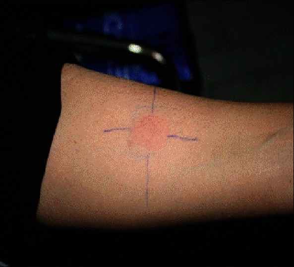
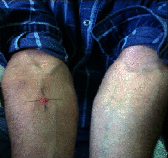
This was a descriptive cross-sectional study. The patients enrolled in this study had three clinical forms of CL confirmed by positive smear of their lesions and then LST was performed for them. Results were categorized as negative (0-5 mm induration), positive (6-14 mm), and strongly positive (≥15 mm). The data were documented in the patients' files and analyzed with SPSS windows software version 16 (Inc.Chicago, USA). RESULTS: 200 patients were enrolled in the study. In the group with plaque type, 86% had a positive LST, 13.3% were negative, and 0.7% were strongly positive. In the lupoid group, these figures were 45.8%, 8.4%, 45.8%, respectively. In the sporotrichoid group, LST was positive in 27.3%, negative in 72.7%, and none of the patients had a strongly positive reaction (P < 0.05). DISCUSSION: The most of the positive LST were belong to plaque and lupoid groups, the most of strongly positive were belong to lupoid, and the most of negative LST were related with sporotrichoid type. CONCLUSION: It can be suggested that lupoid and sporotrichoid types of CL are parts of a continuous spectrum of the disease with an enhanced cellular immunity in lupoid form and a decreased state in sporotrichoid type.

摘要

引言:皮肤利什曼病(CL)是一种具有不同临床形式的寄生虫病。本研究的目的是比较三种形式的皮肤利什曼病(包括斑块型、狼疮样型和孢子丝菌病样型)对利什曼原虫皮肤试验(LST)的反应。 材料与方法:这是一项描述性横断面研究。本研究纳入的患者有三种经病变涂片阳性确诊的皮肤利什曼病临床形式,然后对他们进行利什曼原虫皮肤试验。结果分为阴性(硬结0 - 5毫米)、阳性(6 - 14毫米)和强阳性(≥15毫米)。数据记录在患者档案中,并用SPSS 16版视窗软件(美国芝加哥公司)进行分析。 结果:200名患者纳入本研究。在斑块型组中,86%的患者利什曼原虫皮肤试验呈阳性,13.3%为阴性,0.7%为强阳性。在狼疮样组中,这些数字分别为45.8%、8.4%、45.8%。在孢子丝菌病样组中,利什曼原虫皮肤试验阳性率为27.3%,阴性率为72.7%,且无患者呈强阳性反应(P < 0.05)。 讨论:大多数利什曼原虫皮肤试验阳性的患者属于斑块型和狼疮样组,大多数强阳性患者属于狼疮样组,而大多数利什曼原虫皮肤试验阴性的患者与孢子丝菌病样型有关。 结论:可以认为,狼疮样型和孢子丝菌病样型的皮肤利什曼病是该疾病连续谱的一部分,狼疮样型具有增强的细胞免疫,而孢子丝菌病样型则处于细胞免疫降低状态。

https://cdn.ncbi.nlm.nih.gov/pmc/blobs/467b/3667292/d6d9bd192975/IJD-58-239a-g006.jpg
https://cdn.ncbi.nlm.nih.gov/pmc/blobs/467b/3667292/5f5a1e03c451/IJD-58-239a-g001.jpg
https://cdn.ncbi.nlm.nih.gov/pmc/blobs/467b/3667292/9b9ac697dd0a/IJD-58-239a-g002.jpg
https://cdn.ncbi.nlm.nih.gov/pmc/blobs/467b/3667292/1b9260c71386/IJD-58-239a-g003.jpg
https://cdn.ncbi.nlm.nih.gov/pmc/blobs/467b/3667292/c779614a8b9a/IJD-58-239a-g005.jpg
https://cdn.ncbi.nlm.nih.gov/pmc/blobs/467b/3667292/d6d9bd192975/IJD-58-239a-g006.jpg

相似文献

[1]
Evaluation of leishmanin skin test reaction in different variants of cutaneous leishmaniasis.

Indian J Dermatol. 2013-5

[2]
Evaluation of leishmanin skin test and its relationship with the clinical form and duration of cutaneous leishmaniasis.

Dermatol Online J. 2006-12-10

[3]
Comparison of leishmanin skin test and direct smear for the diagnosis of cutaneous leishmaniasis.

Acta Med Iran. 2011

[4]
Sensitivity of leishmanin skin test in patients of acute cutaneous leishmaniasis.

Dermatol Online J. 2006-5-30

[5]
Comparison of oral itraconazole and intramuscular meglumine antimoniate in the treatment of cutaneous leishmaniasis.

J Coll Physicians Surg Pak. 2007-12

[6]
A review of the leishmanin skin test: A neglected test for a neglected disease.

PLoS Negl Trop Dis. 2021-7

[7]
Surveillance for Leishmania asymptomatic infection in endemic foci of cutaneous leishmaniasis in Venezuela: a combination of leishmanin skin test and PCR using blood clots improves detection and enables identification of species.

Trans R Soc Trop Med Hyg. 2020-6-1

[8]
Atypical and unusual morphological variants of cutaneous leishmaniasis.

Int J Clin Pract. 2021-3

[9]
Immunopathology of anthroponotic cutaneous leishmaniasis and incidental diagnostic tool of metastatic granuloma: A case-control study.

Microb Pathog. 2021-3

[10]
Impaired Th1 Response Is Associated With Therapeutic Failure in Patients With Cutaneous Leishmaniasis Caused by Leishmania braziliensis.

J Infect Dis. 2021-2-13

引用本文的文献

[1]
Comparative Analysis of Microscopy, Leishmanin Skin Test (LST), and Direct Agglutination Test (DAT) for the Diagnosis of Human Cutaneous Leishmaniasis.

Iran J Parasitol. 2025

[2]
A Prospective cohort study of zoonotic cutaneous leishmaniasis in tunisia: Clinical and Immunological features and immune correlates of protection.

PLoS Negl Trop Dis. 2023-12

[3]
Potential biomarkers of immune protection in human leishmaniasis.

Med Microbiol Immunol. 2021-6

[4]
An epidemiological study of visceral leishmaniasis in North East Ethiopia using serological and leishmanin skin tests.

PLoS One. 2019-12-27

[5]
Prevalence of Cutaneous Leishmaniasis in Western Highlands in Yemen.

J Trop Med. 2019-2-28

[6]
Biological evaluation of mimetic peptides as active molecules for a new and simple skin test in an animal model.

Parasitol Res. 2019-1

[7]
Epidemiology of visceral leishmaniasis in Georgia.

PLoS Negl Trop Dis. 2014-3-6

本文引用的文献

[1]
[Sporotrichoid cutaneous leishmaniasis in Tunisia: a clinical and histological study].

Ann Dermatol Venereol. 2008-1

[2]
Cell-mediated immunity in nasopharyngeal carcinoma and allergic rhinitis: a controlled study.

Ear Nose Throat J. 2007-5

[3]
Sensitivity of leishmanin skin test in patients of acute cutaneous leishmaniasis.

Dermatol Online J. 2006-5-30

[4]
Immuno-biochemical evaluations of phenol and thimerosal as antigen preservatives in Montenegro skin test.

Acta Trop. 2006-4

[5]
Atypical clinical variants in New World cutaneous leishmaniasis: disseminated, erysipeloid, and recidiva cutis due to Leishmania (V.) panamensis.

Am J Trop Med Hyg. 2005-8

[6]
Value of diagnostic techniques for cutaneous leishmaniasis.

J Am Acad Dermatol. 2003-7

[7]
Cutaneous leishmaniasis: clinical features and diagnosis.

Clin Dermatol. 1999

[8]
Montenegro skin test--evaluation of the composition and stability of the antigen preparation.

Mem Inst Oswaldo Cruz. 1996

[9]
Evaluation of the efficacy of Iran leishmanin and comparison with leishmanins from Wellcome (UK) and Roma (Italy) in cured cutaneous leishmaniasis patients.

Trans R Soc Trop Med Hyg. 1993

[10]
Diffuse cutaneous leishmaniasis in Ethiopia. I. The clinical and histological features of the disease.

Trans R Soc Trop Med Hyg. 1969

文献AI研究员

20分钟写一篇综述,助力文献阅读效率提升50倍

立即体验

用中文搜PubMed

大模型驱动的PubMed中文搜索引擎

马上搜索

推荐工具

医学文档翻译智能文献检索