• 文献检索
  • 文档翻译
  • 深度研究
  • 学术资讯
  • Suppr Zotero 插件Zotero 插件
  • 邀请有礼
  • 套餐&价格
  • 历史记录
应用&插件
Suppr Zotero 插件Zotero 插件浏览器插件Mac 客户端Windows 客户端微信小程序
定价
高级版会员购买积分包购买API积分包
服务
文献检索文档翻译深度研究API 文档MCP 服务
关于我们
关于 Suppr公司介绍联系我们用户协议隐私条款
关注我们

Suppr 超能文献

核心技术专利:CN118964589B侵权必究
粤ICP备2023148730 号-1Suppr @ 2026

文献检索

告别复杂PubMed语法,用中文像聊天一样搜索,搜遍4000万医学文献。AI智能推荐,让科研检索更轻松。

立即免费搜索

文件翻译

保留排版,准确专业,支持PDF/Word/PPT等文件格式,支持 12+语言互译。

免费翻译文档

深度研究

AI帮你快速写综述,25分钟生成高质量综述,智能提取关键信息,辅助科研写作。

立即免费体验

3D 图像配准对于通过体内微计算机断层扫描(μCT)确保准确检测大鼠胫骨的小梁骨密度、微结构和刚度测量的纵向变化至关重要。

3D image registration is critical to ensure accurate detection of longitudinal changes in trabecular bone density, microstructure, and stiffness measurements in rat tibiae by in vivo microcomputed tomography (μCT).

机构信息

McKay Orthopaedic Research Laboratory, Department of Orthopaedic Surgery, Perelman School of Medicine, University of Pennsylvania, Philadelphia, PA 19104, USA.

出版信息

Bone. 2013 Sep;56(1):83-90. doi: 10.1016/j.bone.2013.05.014. Epub 2013 May 28.

DOI:10.1016/j.bone.2013.05.014
PMID:23727434
原文链接:https://pmc.ncbi.nlm.nih.gov/articles/PMC3715966/
Abstract

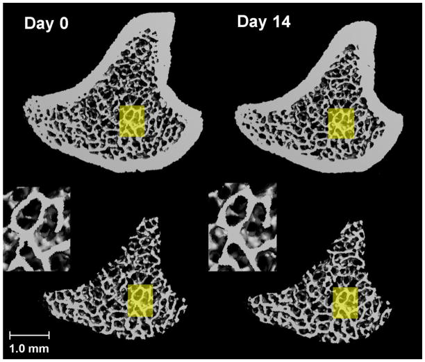
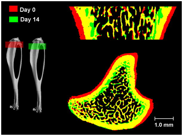
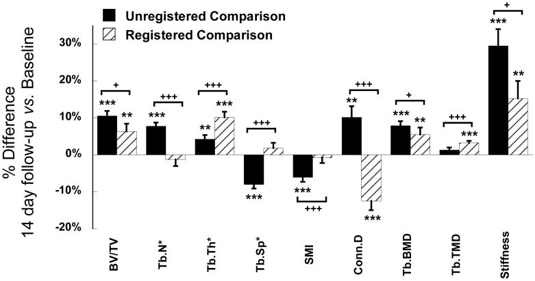
In the recent decade, in vivo μCT scanners have become available to monitor temporal changes in rodent bone in response to diseases and treatments. We investigated short-term and long-term precision of in vivo μCT measurements of trabecular bone density, microstructure and stiffness of rat tibiae and tested whether they can be improved by 3D image registration. Rats in the short-term precision group underwent baseline and follow-up scans within the same day (n = 15) and those in the long-term precision group were scanned at day 0 and day 14 (n = 16) at 10.5 μm voxel size. A 3D image-registration scheme was applied to register the trabecular bone compartments of baseline and follow-up scans. Prior to image registration, short-term precision ranged between 0.85% and 2.65% in bone volume fraction (BV/TV), trabecular number, thickness, and spacing (Tb.N*, Tb.Th*, Tb.Sp*), trabecular bone mineral density and tissue mineral density (Tb.BMD, and Tb.TMD), and was particularly high in structure model index (SMI), connectivity density (Conn.D), and stiffness (4.29%-8.83%). Image registration tended to improve the short-term precision, but the only statistically significant improvement was in Tb.N*, Tb.TMD, and stiffness. On the other hand, unregistered comparisons between day-0 and day-14 scans suggested significant increases in BV/TV, Tb.N*, Tb.Th*, Conn.D, and Tb.BMD and decrease in Tb.Sp* and SMI. However, the percent change in each parameter from registered comparisons was significantly different from unregistered comparisons. Registered results suggested a significant increase in BV/TV, Tb.BMD, and stiffness over 14 days, primarily caused by increased Tb.Th* and Tb.TMD. Due to the continuous growth of rodents, the direct comparisons between the unregistered baseline and follow-up scans were driven by changes due to global bone modeling instead of local remodeling. Our results suggested that 3D image registration is critical for detecting changes due to bone remodeling activities in rodent trabecular bone by in vivo μCT imaging.

摘要

在最近的十年中,活体 μCT 扫描仪已经可以用于监测疾病和治疗对啮齿动物骨骼的时间变化。我们研究了活体 μCT 测量胫骨小梁骨密度、微结构和刚度的短期和长期精度,并测试了 3D 图像配准是否可以提高这些精度。短期精度组的大鼠在同一天内进行基线和随访扫描(n=15),而长期精度组的大鼠在第 0 天和第 14 天(n=16)以 10.5μm 体素大小进行扫描。应用 3D 图像配准方案对基线和随访扫描的小梁骨区进行配准。在图像配准之前,短期精度范围在骨体积分数(BV/TV)、小梁数量、厚度和间隔(Tb.N*、Tb.Th*、Tb.Sp*)、小梁骨密度和组织骨密度(Tb.BMD 和 Tb.TMD)的 0.85%-2.65%之间,结构模型指数(SMI)、连通密度(Conn.D)和刚度(4.29%-8.83%)的精度尤其高。图像配准往往会提高短期精度,但唯一具有统计学意义的提高是在 Tb.N*、Tb.TMD 和刚度方面。另一方面,未配准的第 0 天和第 14 天扫描之间的比较表明,BV/TV、Tb.N*、Tb.Th*、Conn.D 和 Tb.BMD 显著增加,而 Tb.Sp和 SMI 则减少。然而,从已配准的比较中得到的每个参数的百分比变化与未配准的比较明显不同。已配准的结果表明,14 天内 BV/TV、Tb.BMD 和刚度显著增加,主要是由于 Tb.Th和 Tb.TMD 的增加。由于啮齿动物的持续生长,未配准的基线和随访扫描之间的直接比较是由全局骨建模引起的变化驱动的,而不是由局部重塑引起的变化。我们的结果表明,3D 图像配准对于通过活体 μCT 成像检测啮齿动物小梁骨中由于骨重塑活动引起的变化至关重要。

https://cdn.ncbi.nlm.nih.gov/pmc/blobs/952a/3715966/fd34740292d1/nihms485775f7.jpg
https://cdn.ncbi.nlm.nih.gov/pmc/blobs/952a/3715966/2973ecc2cde8/nihms485775f1.jpg
https://cdn.ncbi.nlm.nih.gov/pmc/blobs/952a/3715966/6b67e5fb7bef/nihms485775f2.jpg
https://cdn.ncbi.nlm.nih.gov/pmc/blobs/952a/3715966/3aad4782985b/nihms485775f3.jpg
https://cdn.ncbi.nlm.nih.gov/pmc/blobs/952a/3715966/13d3b77b67ba/nihms485775f4.jpg
https://cdn.ncbi.nlm.nih.gov/pmc/blobs/952a/3715966/017fffb44c9f/nihms485775f5.jpg
https://cdn.ncbi.nlm.nih.gov/pmc/blobs/952a/3715966/760c1181b367/nihms485775f6.jpg
https://cdn.ncbi.nlm.nih.gov/pmc/blobs/952a/3715966/fd34740292d1/nihms485775f7.jpg

相似文献

1
3D image registration is critical to ensure accurate detection of longitudinal changes in trabecular bone density, microstructure, and stiffness measurements in rat tibiae by in vivo microcomputed tomography (μCT).3D 图像配准对于通过体内微计算机断层扫描(μCT)确保准确检测大鼠胫骨的小梁骨密度、微结构和刚度测量的纵向变化至关重要。
Bone. 2013 Sep;56(1):83-90. doi: 10.1016/j.bone.2013.05.014. Epub 2013 May 28.
2
High-resolution peripheral quantitative computed tomography can assess microstructural and mechanical properties of human distal tibial bone.高分辨率外周定量计算机断层扫描可评估人类胫骨远端骨的微观结构和力学性能。
J Bone Miner Res. 2010 Apr;25(4):746-56. doi: 10.1359/jbmr.090822.
3
Analysis of Bone Histomorphometry in Rat and Guinea Pig Animal Models Subject to Hypoxia.缺氧状态下大鼠和豚鼠动物模型的骨组织形态计量学分析。
Int J Mol Sci. 2022 Oct 22;23(21):12742. doi: 10.3390/ijms232112742.
4
Bone quality in patients with osteoporosis undergoing lumbar fusion surgery: analysis of the MRI-based vertebral bone quality score and the bone microstructure derived from microcomputed tomography.骨质疏松症患者行腰椎融合术的骨质量:基于 MRI 的椎体骨质量评分和来自微计算机断层扫描的骨微观结构分析。
Spine J. 2022 Oct;22(10):1642-1650. doi: 10.1016/j.spinee.2022.05.008. Epub 2022 Jun 5.
5
High-resolution peripheral quantitative computed tomography (HR-pQCT) can assess microstructural and biomechanical properties of both human distal radius and tibia: Ex vivo computational and experimental validations.高分辨率外周定量计算机断层扫描(HR-pQCT)可评估人体桡骨远端和胫骨的微观结构及生物力学特性:离体计算与实验验证。
Bone. 2016 May;86:58-67. doi: 10.1016/j.bone.2016.02.016. Epub 2016 Feb 26.
6
Accuracy of trabecular structure by HR-pQCT compared to gold standard μCT in the radius and tibia of patients with osteoporosis and long-term bisphosphonate therapy.在接受长期双膦酸盐治疗的骨质疏松症患者的桡骨和胫骨中,与金标准微计算机断层扫描(μCT)相比,高分辨率外周定量计算机断层扫描(HR-pQCT)对小梁结构的准确性。
Osteoporos Int. 2014 May;25(5):1595-606. doi: 10.1007/s00198-014-2650-4. Epub 2014 Feb 25.
7
[Analysis of correlation between trabecular microstructure and clinical imaging parameters in fracture region of osteoporotic hip].[骨质疏松性髋部骨折区域小梁微结构与临床影像参数的相关性分析]
Zhongguo Xiu Fu Chong Jian Wai Ke Za Zhi. 2014 May;28(5):576-80.
8
Microcomputed tomography analysis of particular autogenous bone graft in sinus augmentation at 5 months: differences on bone mineral density and 3D trabecular structure.5 个月时鼻窦提升中特定自体骨移植物的微计算机断层分析:骨密度和 3D 小梁结构的差异。
Clin Oral Investig. 2013 Mar;17(2):535-42. doi: 10.1007/s00784-012-0725-1. Epub 2012 Apr 25.
9
Validation of calcaneus trabecular microstructure measurements by HR-pQCT.跟骨小梁微结构测量的 HR-pQCT 验证。
Bone. 2018 Jan;106:69-77. doi: 10.1016/j.bone.2017.09.013. Epub 2017 Oct 3.
10
Quantification of bone microstructure in the wrist using cone-beam computed tomography.利用锥形束 CT 对腕骨骨微结构进行定量分析。
Bone. 2018 Sep;114:206-214. doi: 10.1016/j.bone.2018.06.006. Epub 2018 Jun 15.

引用本文的文献

1
Anti-Siglec-15 Antibody Prevents Marked Bone Loss after Acute Spinal Cord Injury-Induced Immobilization in Rats.抗唾液酸结合免疫球蛋白样凝集素15抗体可预防大鼠急性脊髓损伤诱导制动后的明显骨质流失。
JBMR Plus. 2023 Sep 27;7(12):e10825. doi: 10.1002/jbm4.10825. eCollection 2023 Dec.
2
Distinct Responses of Modeling- and Remodeling-Based Bone Formation to the Discontinuation of Intermittent Parathyroid Hormone Treatment in Ovariectomized Rats.间断甲状旁腺激素治疗对去卵巢大鼠模型和重塑型骨形成的不同反应。
J Bone Miner Res. 2022 Nov;37(11):2215-2225. doi: 10.1002/jbmr.4704. Epub 2022 Sep 30.
3
Administration of High-Dose Methylprednisolone Worsens Bone Loss after Acute Spinal Cord Injury in Rats.

本文引用的文献

1
Mouse tail vertebrae adapt to cyclic mechanical loading by increasing bone formation rate and decreasing bone resorption rate as shown by time-lapsed in vivo imaging of dynamic bone morphometry.动态骨形态计量学的实时活体成像显示,小鼠尾椎通过增加成骨速率和降低骨吸收速率来适应循环机械载荷。
Bone. 2011 Dec;49(6):1340-50. doi: 10.1016/j.bone.2011.08.035. Epub 2011 Sep 18.
2
MicroCT morphometry analysis of mouse cancellous bone: intra- and inter-system reproducibility.小鼠松质骨的 microCT 形态计量分析:内系统和外系统的可重复性。
Bone. 2011 Sep;49(3):580-7. doi: 10.1016/j.bone.2011.05.013. Epub 2011 May 20.
3
In vivo micro-computed tomography allows direct three-dimensional quantification of both bone formation and bone resorption parameters using time-lapsed imaging.
大剂量甲基强的松龙给药会加重大鼠急性脊髓损伤后的骨质流失。
Neurotrauma Rep. 2021 Dec 8;2(1):592-602. doi: 10.1089/neur.2021.0035. eCollection 2021.
4
Short Cyclic Regimen With Parathyroid Hormone (PTH) Results in Prolonged Anabolic Effect Relative to Continuous Treatment Followed by Discontinuation in Ovariectomized Rats.与连续给予甲状旁腺激素(PTH)然后停药相比,在去卵巢大鼠中采用甲状旁腺激素短周期治疗方案可产生更持久的合成代谢效应。
J Bone Miner Res. 2022 Apr;37(4):616-628. doi: 10.1002/jbmr.4495. Epub 2022 Jan 20.
5
Activation, development, and attenuation of modeling- and remodeling-based bone formation in adult rats.成年大鼠基于建模和重塑的骨形成的激活、发育和衰减。
Biomaterials. 2021 Sep;276:121015. doi: 10.1016/j.biomaterials.2021.121015. Epub 2021 Jul 9.
6
Maternal bone adaptation to mechanical loading during pregnancy, lactation, and post-weaning recovery.母体骨骼对妊娠、哺乳期和离乳后恢复期机械负荷的适应。
Bone. 2021 Oct;151:116031. doi: 10.1016/j.bone.2021.116031. Epub 2021 Jun 5.
7
Peak trabecular bone microstructure predicts rate of estrogen-deficiency-induced bone loss in rats.峰值骨小梁微结构可预测大鼠雌激素缺乏诱导的骨丢失率。
Bone. 2021 Apr;145:115862. doi: 10.1016/j.bone.2021.115862. Epub 2021 Jan 22.
8
Electrical stimulation of hindlimb skeletal muscle has beneficial effects on sublesional bone in a rat model of spinal cord injury.电刺激后肢骨骼肌对脊髓损伤大鼠模型的亚损伤骨具有有益作用。
Bone. 2021 Mar;144:115825. doi: 10.1016/j.bone.2020.115825. Epub 2020 Dec 19.
9
Shear Stress in Bone Marrow has a Dose Dependent Effect on cFos Gene Expression in Culture.骨髓中的剪切应力对培养中的cFos基因表达具有剂量依赖性效应。
Cell Mol Bioeng. 2019 Aug 27;12(6):559-568. doi: 10.1007/s12195-019-00594-z. eCollection 2019 Dec.
10
Rapid bone loss occurs as early as 2 days after complete spinal cord transection in young adult rats.在年轻成年大鼠中,完全性脊髓横断后仅 2 天就会发生快速骨丢失。
Spinal Cord. 2020 Mar;58(3):309-317. doi: 10.1038/s41393-019-0371-4. Epub 2019 Oct 29.
体内 micro-CT 允许使用时变成像直接对骨形成和骨吸收参数进行三维定量。
Bone. 2011 Mar 1;48(3):433-42. doi: 10.1016/j.bone.2010.10.007. Epub 2010 Oct 13.
4
Abnormal microarchitecture and reduced stiffness at the radius and tibia in postmenopausal women with fractures.绝经后骨折妇女桡骨和胫骨微观结构异常和刚度降低。
J Bone Miner Res. 2010 Dec;25(12):2572-81. doi: 10.1002/jbmr.152. Epub 2010 Jun 18.
5
Guidelines for assessment of bone microstructure in rodents using micro-computed tomography.骨组织形态计量学分析的鼠类骨骼microCT 评估指南
J Bone Miner Res. 2010 Jul;25(7):1468-86. doi: 10.1002/jbmr.141.
6
Finite element analysis performed on radius and tibia HR-pQCT images and fragility fractures at all sites in postmenopausal women.对绝经后妇女桡骨和胫骨 HR-pQCT 图像及所有部位脆性骨折进行有限元分析。
Bone. 2010 Apr;46(4):1030-7. doi: 10.1016/j.bone.2009.12.015. Epub 2010 Jan 4.
7
Reproducibility of bone micro-architecture measurements in rodents by in vivo micro-computed tomography is maximized with three-dimensional image registration.在体小动物 micro-CT 骨微结构测量的可重复性可以通过三维图像配准来实现。
Bone. 2010 Jan;46(1):155-61. doi: 10.1016/j.bone.2009.09.023. Epub 2009 Sep 29.
8
High-resolution peripheral quantitative computed tomography can assess microstructural and mechanical properties of human distal tibial bone.高分辨率外周定量计算机断层扫描可评估人类胫骨远端骨的微观结构和力学性能。
J Bone Miner Res. 2010 Apr;25(4):746-56. doi: 10.1359/jbmr.090822.
9
Assessing forearm fracture risk in postmenopausal women.评估绝经后妇女的前臂骨折风险。
Osteoporos Int. 2010 Jul;21(7):1161-9. doi: 10.1007/s00198-009-1047-2. Epub 2009 Aug 28.
10
Assessment of trabecular bone microarchitecture by two different x-ray microcomputed tomographs: a comparative study of the rat distal tibia using Skyscan and Scanco devices.使用两种不同的X射线显微计算机断层扫描仪评估小梁骨微结构:使用Skyscan和Scanco设备对大鼠胫骨远端进行的比较研究。
Med Phys. 2009 Apr;36(4):1286-97. doi: 10.1118/1.3096605.