• 文献检索
  • 文档翻译
  • 深度研究
  • 学术资讯
  • Suppr Zotero 插件Zotero 插件
  • 邀请有礼
  • 套餐&价格
  • 历史记录
应用&插件
Suppr Zotero 插件Zotero 插件浏览器插件Mac 客户端Windows 客户端微信小程序
定价
高级版会员购买积分包购买API积分包
服务
文献检索文档翻译深度研究API 文档MCP 服务
关于我们
关于 Suppr公司介绍联系我们用户协议隐私条款
关注我们

Suppr 超能文献

核心技术专利:CN118964589B侵权必究
粤ICP备2023148730 号-1Suppr @ 2026

文献检索

告别复杂PubMed语法,用中文像聊天一样搜索,搜遍4000万医学文献。AI智能推荐,让科研检索更轻松。

立即免费搜索

文件翻译

保留排版,准确专业,支持PDF/Word/PPT等文件格式,支持 12+语言互译。

免费翻译文档

深度研究

AI帮你快速写综述,25分钟生成高质量综述,智能提取关键信息,辅助科研写作。

立即免费体验

手动、数字和侧位 CBCT 头影测量分析的比较。

Comparison of manual, digital and lateral CBCT cephalometric analyses.

机构信息

Oral and Maxillofacial Surgery Division, Saint Francis Hospital, Cambé, PR, Brazil.

出版信息

J Appl Oral Sci. 2013 Mar-Apr;21(2):167-76. doi: 10.1590/1678-7757201302326.

DOI:10.1590/1678-7757201302326
PMID:23739848
原文链接:https://pmc.ncbi.nlm.nih.gov/articles/PMC3881882/
Abstract

OBJECTIVE

The aim of this study was to compare the reliability of three different methods of cephalometric analysis.

MATERIAL AND METHODS

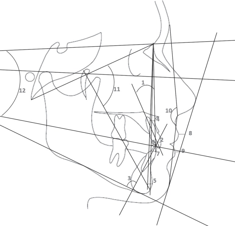
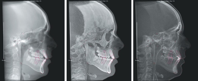
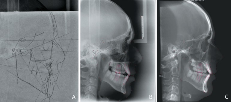
Conventional pretreatment lateral cephalograms and cone beam computed tomography (CBCT) scans from 50 subjects from a radiological clinic were selected in order to test the three methods: manual tracings (MT), digitized lateral cephalograms (DLC), and lateral cephalograms from CBCT (LC-CBCT). The lateral cephalograms were manually analyzed through the Dolphin Imaging 11.0™ software. Twenty measurements were performed under the same conditions, and retraced after a 30-day period. Paired t tests and the Dahlberg formula were used to evaluate the intra-examiner errors. The Pearson's correlation coefficient and one-way analysis of variance (ANOVA) tests were used to compare the differences between the methods.

RESULTS

Intra-examiner reliability occurred for all methods for most of the measurements. Only six measurements were different between the methods and an agreement was observed in the analyses among the 3 methods.

CONCLUSIONS

The results demonstrated that all evaluated methodologies are reliable and valid for scientific research, however, the method used in the lateral cephalograms from the CBCT proved the most reliable.

摘要

目的

本研究旨在比较三种不同的头影测量分析方法的可靠性。

材料与方法

从一家放射科诊所选择了 50 名患者的常规预处理侧位头颅 X 光片和锥形束 CT(CBCT)扫描,以测试三种方法:手动描记(MT)、数字化侧位头颅 X 光片(DLC)和 CBCT 的侧位头颅 X 光片(LC-CBCT)。使用 Dolphin Imaging 11.0™ 软件对手动侧位头颅 X 光片进行分析。在相同条件下进行了 20 次测量,并在 30 天后进行了重新测量。使用配对 t 检验和 Dahlberg 公式评估内部检验员误差。使用 Pearson 相关系数和单向方差分析(ANOVA)检验来比较方法之间的差异。

结果

所有方法对于大多数测量的内部检验员可靠性均得到验证。只有 6 项测量值在方法之间存在差异,并且在 3 种方法的分析中观察到了一致性。

结论

结果表明,所有评估的方法对于科学研究都是可靠和有效的,但是,来自 CBCT 的侧位头颅 X 光片中使用的方法被证明最可靠。

https://cdn.ncbi.nlm.nih.gov/pmc/blobs/65da/3881882/fe21f7eb94ad/jaos-21-02-0167-g03.jpg
https://cdn.ncbi.nlm.nih.gov/pmc/blobs/65da/3881882/1e2d8d459cdb/jaos-21-02-0167-g01.jpg
https://cdn.ncbi.nlm.nih.gov/pmc/blobs/65da/3881882/f7a46f95ffd1/jaos-21-02-0167-g02.jpg
https://cdn.ncbi.nlm.nih.gov/pmc/blobs/65da/3881882/fe21f7eb94ad/jaos-21-02-0167-g03.jpg
https://cdn.ncbi.nlm.nih.gov/pmc/blobs/65da/3881882/1e2d8d459cdb/jaos-21-02-0167-g01.jpg
https://cdn.ncbi.nlm.nih.gov/pmc/blobs/65da/3881882/f7a46f95ffd1/jaos-21-02-0167-g02.jpg
https://cdn.ncbi.nlm.nih.gov/pmc/blobs/65da/3881882/fe21f7eb94ad/jaos-21-02-0167-g03.jpg

相似文献

1
Comparison of manual, digital and lateral CBCT cephalometric analyses.手动、数字和侧位 CBCT 头影测量分析的比较。
J Appl Oral Sci. 2013 Mar-Apr;21(2):167-76. doi: 10.1590/1678-7757201302326.
2
The reliability of cephalometric measurements in oral and maxillofacial imaging: Cone beam computed tomography versus two-dimensional digital cephalograms.口腔颌面影像学中头影测量的可靠性:锥形束计算机断层扫描与二维数字化头颅侧位片的比较
Indian J Dent Res. 2016 Jul-Aug;27(4):370-377. doi: 10.4103/0970-9290.191884.
3
Accuracy of linear measurements from imaging plate and lateral cephalometric images derived from cone-beam computed tomography.成像板及锥形束计算机断层扫描所得头颅侧位影像的线性测量准确性
Am J Orthod Dentofacial Orthop. 2007 Oct;132(4):550-60. doi: 10.1016/j.ajodo.2006.09.046.
4
A comparative analysis of angular cephalometric values between CBCT generated lateral cephalograms versus digitized conventional lateral cephalograms.CBCT生成的头颅侧位片与数字化传统头颅侧位片之间的角度头影测量值的比较分析。
Int Orthod. 2009 Dec;7(4):308-21. doi: 10.1016/S1761-7227(09)73505-3. Epub 2010 Jan 30.
5
Comparison of linear and angular measurements using two-dimensional conventional methods and three-dimensional cone beam CT images reconstructed from a volumetric rendering program in vivo.体内使用二维传统方法和从容积再现程序重建的三维锥形束 CT 图像进行线性和角度测量的比较。
Dentomaxillofac Radiol. 2011 Dec;40(8):492-500. doi: 10.1259/dmfr/15644321.
6
Comparison between conventional and cone-beam computed tomography-generated cephalograms.传统头颅侧位片与锥形束计算机断层扫描生成的头颅侧位片的比较。
Am J Orthod Dentofacial Orthop. 2008 Dec;134(6):798-802. doi: 10.1016/j.ajodo.2008.07.008.
7
Reliability of cephalograms derived of cone beam computed tomography versus lateral cephalograms to estimate cervical vertebrae maturity in a Peruvian population: A retrospective study.基于锥形束 CT 的头颅侧位片与传统头颅侧位片评估秘鲁人群颈椎成熟度的可靠性:一项回顾性研究。
Int Orthod. 2020 Jun;18(2):258-265. doi: 10.1016/j.ortho.2020.01.001. Epub 2020 Jan 31.
8
Cephalometric measurements performed on CBCT and reconstructed lateral cephalograms: a cross-sectional study providing a quantitative approach of differences and bias.CBCT 上进行的头影测量和重建的侧位头颅侧位片:提供定量分析差异和偏差的横断面研究。
BMC Oral Health. 2022 Mar 30;22(1):98. doi: 10.1186/s12903-022-02131-3.
9
Accuracy of linear measurements using three imaging modalities: two lateral cephalograms and one 3D model from CBCT data.使用三种成像方式进行线性测量的准确性:两张侧位头影测量片和一张来自锥形束CT数据的三维模型。
Eur J Orthod. 2015 Apr;37(2):202-8. doi: 10.1093/ejo/cju036. Epub 2014 Aug 26.
10
Cervical vertebral column morphology in patients with obstructive sleep apnoea assessed using lateral cephalograms and cone beam CT. A comparative study.采用侧位头颅片和锥形束 CT 评估阻塞性睡眠呼吸暂停患者的颈椎柱形态。一项对比研究。
Dentomaxillofac Radiol. 2013;42(6):20130060. doi: 10.1259/dmfr.20130060. Epub 2013 Mar 15.

引用本文的文献

1
Accuracy and Reproducibility of Semidigital Versus Fully Digital Cephalometric Tracings Using a New Computer Program Versus Conventional Methods (Gold Standards): A Preliminary Study.使用新计算机程序与传统方法(金标准)对比半数字与全数字头影测量描记的准确性和可重复性:一项初步研究。
Biomed Res Int. 2025 Aug 18;2025:8403357. doi: 10.1155/bmri/8403357. eCollection 2025.
2
Cone beam computed tomography analysis of anterior open bite management using clear aligners: a single-arm retrospective study.使用透明矫治器治疗前牙开(牙合)的锥形束计算机断层扫描分析:一项单臂回顾性研究。
Sci Rep. 2025 Jul 11;15(1):25145. doi: 10.1038/s41598-025-09157-x.
3

本文引用的文献

1
Reliability of a method to conduct upper airway analysis in cone-beam computed tomography.一种在锥形束计算机断层扫描中进行上气道分析的方法的可靠性。
Braz Oral Res. 2013 Jan-Feb;27(1):48-54. doi: 10.1590/s1806-83242013000100009.
2
Reliability and the smallest detectable difference of measurements on 3-dimensional cone-beam computed tomography images.三维锥形束计算机断层摄影图像测量的可靠性和最小可检测差值。
Am J Orthod Dentofacial Orthop. 2011 Sep;140(3):e107-14. doi: 10.1016/j.ajodo.2011.02.020.
3
Craniofacial characteristics of Caucasian and Afro-Caucasian Brazilian subjects with normal occlusion.
Inclination Changes in Incisors During Orthodontic Treatment with Passive Self-Ligating Brackets.
使用被动自结扎托槽进行正畸治疗期间切牙的倾斜度变化
J Clin Med. 2025 May 12;14(10):3370. doi: 10.3390/jcm14103370.
4
Can MRI be a potential substitute for CT in cephalometric analysis for radiation-free diagnoses?在头影测量分析中,磁共振成像(MRI)能否成为计算机断层扫描(CT)的潜在替代品,用于无辐射诊断?
J Orofac Orthop. 2025 Mar 10. doi: 10.1007/s00056-025-00576-z.
5
Digital versus Manual Tracing in Cephalometric Analysis: A Systematic Review and Meta-Analysis.头影测量分析中数字追踪与手工追踪的比较:系统评价与Meta分析
J Pers Med. 2024 May 25;14(6):566. doi: 10.3390/jpm14060566.
6
Comparative Evaluation of Digital Cephalometric Tracing Applications on Mobile Devices and Manual Tracing.移动设备上的数字化头影测量追踪应用与手动追踪的比较评估
Med Sci Monit. 2024 Jun 23;30:e944628. doi: 10.12659/MSM.944628.
7
Comparing the Effect of Miniscrew-Supported and Conventional Maxillary Incisor Intrusion on the Inclination of Maxillary Incisors and Molars - A Systematic Review and Meta-Analysis.比较微型螺钉支持的上颌切牙压低与传统上颌切牙压低对上颌切牙和磨牙倾斜度的影响——一项系统评价和荟萃分析
Contemp Clin Dent. 2022 Oct-Dec;13(4):307-314. doi: 10.4103/ccd.ccd_385_22. Epub 2022 Dec 1.
8
Diagnostic value of routine dental radiographs for predicting the mandibular canal localization validated by cone-beam computed tomogram measurements.常规牙片预测下颌管定位的诊断价值通过锥形束 CT 测量得到验证。
Clin Exp Dent Res. 2022 Dec;8(6):1440-1448. doi: 10.1002/cre2.639. Epub 2022 Aug 8.
9
Informational content of two-dimensional panoramic radiographs and lateral cephalometric radiographs with respect to the bone volume of intraoral donor regions considering CBCT imaging.二维全景片和侧位头颅定位片与 CBCT 成像的口腔供区骨量的信息内容。
BMC Oral Health. 2022 Jul 30;22(1):318. doi: 10.1186/s12903-022-02344-6.
10
Three-Dimensional Analysis of Posterior Mandibular Displacement in Rats.大鼠下颌骨后移位的三维分析
Vet Sci. 2022 Mar 20;9(3):144. doi: 10.3390/vetsci9030144.
正常牙合的白种人和非裔白种巴西人颅面特征。
J Appl Oral Sci. 2011 Apr;19(2):118-24. doi: 10.1590/s1678-77572011000200007.
4
Picture archiving and communications systems: a study of reliability of orthodontic cephalometric analysis.影像归档和通信系统:正畸头影测量分析可靠性的研究。
Eur J Orthod. 2011 Oct;33(5):537-43. doi: 10.1093/ejo/cjq116. Epub 2010 Nov 24.
5
Comparison of common hard tissue cephalometric measurements between computed tomography 3D reconstruction and conventional 2D cephalometric images.比较 CT 三维重建与传统二维头影测量图像常见硬组织测量指标。
Angle Orthod. 2011 Jan;81(1):11-16. doi: 10.2319/031710-157.1.
6
A comparison of skeletal, dentoalveolar and soft tissue characteristics in white and black Brazilian subjects.白人和黑人巴西人群体的骨骼、牙颌和软组织特征比较。
J Appl Oral Sci. 2010 Mar-Apr;18(2):135-42. doi: 10.1590/s1678-77572010000200007.
7
Digital and manual cephalometric analysis.数字化和手工头影测量分析。
Bratisl Lek Listy. 2010;111(2):97-100.
8
Accuracy and landmark error calculation using cone-beam computed tomography-generated cephalograms.使用锥形束计算机断层扫描生成的头颅侧位片计算准确性和标志点误差。
Angle Orthod. 2010 Mar;80(2):286-94. doi: 10.2319/030909-135.1.
9
A comparison between two-dimensional and three-dimensional cephalometry on frontal radiographs and on cone beam computed tomography scans of human skulls.人类颅骨正位X线片和锥形束计算机断层扫描上二维与三维头影测量的比较。
Eur J Oral Sci. 2009 Jun;117(3):300-5. doi: 10.1111/j.1600-0722.2009.00633.x.
10
Differences in cephalometric measurements: a comparison of digital versus hand-tracing methods.头影测量的差异:数字法与手工描绘法的比较
Eur J Orthod. 2009 Jun;31(3):254-9. doi: 10.1093/ejo/cjn121. Epub 2009 Apr 6.