Farahmand P, Marin F, Hawkins F, Möricke R, Ringe J D, Glüer C-C, Papaioannou N, Minisola S, Martínez G, Nolla J M, Niedhart C, Guañabens N, Nuti R, Martín-Mola E, Thomasius F, Peña J, Graeff C, Kapetanos G, Petto H, Gentzel A, Reisinger A, Zysset P K
West German Osteoporosis Centre, Klinikum Leverkusen, University of Cologne, Am Gesundheitspark 11, 51375, Leverkusen, Germany,
Osteoporos Int. 2013 Dec;24(12):2971-81. doi: 10.1007/s00198-013-2379-5. Epub 2013 Jun 6.
Changes of the bone formation marker PINP correlated positively with improvements in vertebral strength in men with glucocorticoid-induced osteoporosis (GIO) who received 18-month treatment with teriparatide, but not with risedronate. These results support the use of PINP as a surrogate marker of bone strength in GIO patients treated with teriparatide.
To investigate the correlations between biochemical markers of bone turnover and vertebral strength estimated by finite element analysis (FEA) in men with GIO.
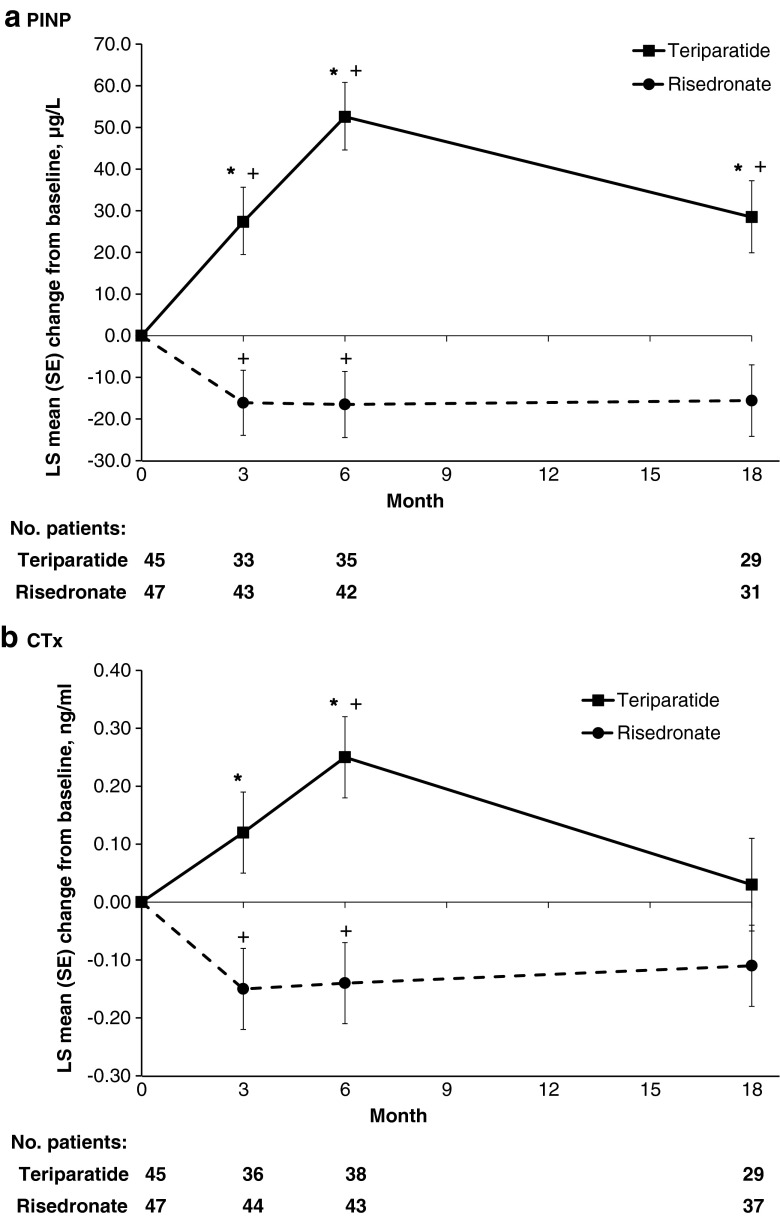
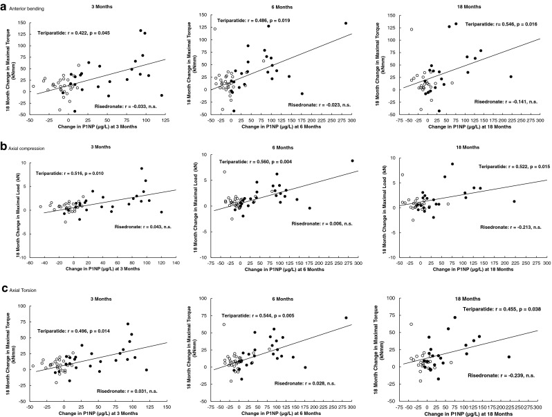
A total of 92 men with GIO were included in an 18-month, randomized, open-label trial of teriparatide (20 μg/day, n = 45) and risedronate (35 mg/week, n = 47). High-resolution quantitative computed tomography images of the 12th thoracic vertebra obtained at baseline, 6 and 18 months were converted into digital nonlinear FE models and subjected to anterior bending, axial compression and torsion. Stiffness and strength were computed for each model and loading mode. Serum biochemical markers of bone formation (amino-terminal-propeptide of type I collagen [PINP]) and bone resorption (type I collagen cross-linked C-telopeptide degradation fragments [CTx]) were measured at baseline, 3 months, 6 months and 18 months. A mixed-model of repeated measures analysed changes from baseline and between-group differences. Spearman correlations assessed the relationship between changes from baseline of bone markers with FEA variables.
PINP and CTx levels increased in the teriparatide group and decreased in the risedronate group. FEA-derived parameters increased in both groups, but were significantly higher at 18 months in the teriparatide group. Significant positive correlations were found between changes from baseline of PINP at 3, 6 and 18 months with changes in FE strength in the teriparatide-treated group, but not in the risedronate group.
Positive correlations between changes in a biochemical marker of bone formation and improvement of biomechanical properties support the use of PINP as a surrogate marker of bone strength in teriparatide-treated GIO patients.
接受18个月特立帕肽治疗的糖皮质激素诱导的骨质疏松症(GIO)男性患者,骨形成标志物I型前胶原氨基端前肽(PINP)的变化与椎体强度的改善呈正相关,但与利塞膦酸盐治疗无关。这些结果支持将PINP用作接受特立帕肽治疗的GIO患者骨强度的替代标志物。
研究GIO男性患者中骨转换生化标志物与通过有限元分析(FEA)估算的椎体强度之间的相关性。
总共92例GIO男性患者纳入了一项为期18个月的特立帕肽(20μg/天,n = 45)和利塞膦酸盐(35mg/周,n = 47)随机、开放标签试验。在基线、6个月和18个月时获得的第12胸椎的高分辨率定量计算机断层扫描图像被转换为数字非线性FE模型,并进行前屈、轴向压缩和扭转。计算每个模型和加载模式的刚度和强度。在基线、3个月、6个月和18个月时测量骨形成(I型胶原氨基端前肽[PINP])和骨吸收(I型胶原交联C末端肽降解片段[CTx])的血清生化标志物。重复测量的混合模型分析了与基线的变化和组间差异。Spearman相关性评估了骨标志物与FEA变量从基线变化之间的关系。
特立帕肽组中PINP和CTx水平升高,利塞膦酸盐组中降低。两组中FEA衍生参数均升高,但特立帕肽组在18个月时显著更高。在特立帕肽治疗组中,3、6和18个月时PINP与基线变化之间与FE强度变化之间存在显著正相关,但在利塞膦酸盐组中不存在。
骨形成生化标志物的变化与生物力学性能改善之间的正相关支持将PINP用作特立帕肽治疗的GIO患者骨强度的替代标志物。