Serra Laura, Cercignani Mara, Carlesimo Giovanni A, Fadda Lucia, Tini Nadia, Giulietti Giovanni, Caltagirone Carlo, Bozzali Marco
Neuroimaging Laboratory, Santa Lucia Foundation, IRCCS, Rome, Italy.
PLoS One. 2013 Jun 3;8(6):e64578. doi: 10.1371/journal.pone.0064578. Print 2014.
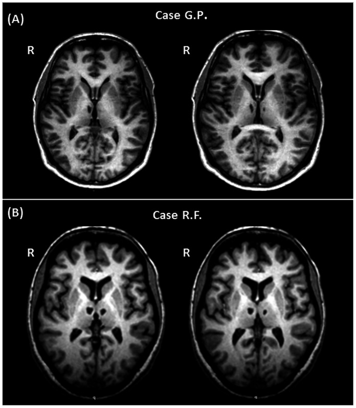
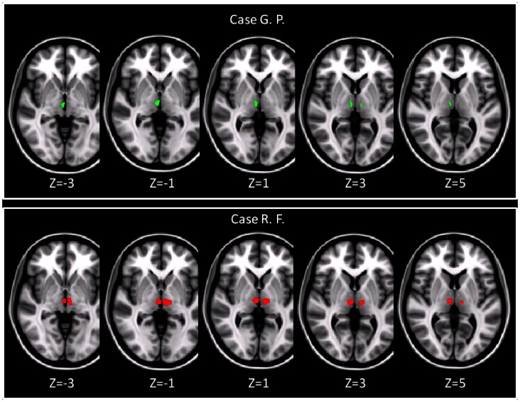
A novel approach based on diffusion tractography was used here to characterise the cortico-thalamic connectivity in two patients, both presenting with an isolated bilateral infarct in the thalamus, but exhibiting partially different cognitive and behavioural profiles. Both patients (G.P. and R.F.) had a pervasive deficit in episodic memory, but only one of them (R.F.) suffered also from a dysexecutive syndrome. Both patients had an MRI scan at 3T, including a T1-weighted volume. Their lesions were manually segmented. T1-volumes were normalised to standard space, and the same transformations were applied to the lesion masks. Nineteen healthy controls underwent a diffusion-tensor imaging (DTI) scan. Their DTI data were normalised to standard space and averaged. An atlas of Brodmann areas was used to parcellate the prefrontal cortex. Probabilistic tractography was used to assess the probability of connection between each voxel of the thalamus and a set of prefrontal areas. The resulting map of corticothalamic connections was superimposed onto the patients' lesion masks, to assess whether the location of the thalamic lesions in R.F. (but not in G. P.) implied connections with prefrontal areas involved in dysexecutive syndromes. In G.P., the lesion fell within areas of the thalamus poorly connected with prefrontal areas, showing only a modest probability of connection with the anterior cingulate cortex (ACC). Conversely, R.F.'s lesion fell within thalamic areas extensively connected with the ACC bilaterally, with the right dorsolateral prefrontal cortex, and with the left supplementary motor area. Despite a similar, bilateral involvement of the thalamus, the use of connectivity-based segmentation clarified that R.F.'s lesions only were located within nuclei highly connected with the prefrontal cortical areas, thus explaining the patient's frontal syndrome. This study confirms that DTI tractography is a useful tool to examine in vivo the effect of focal lesions on interconnectivity brain patterns.
本文采用了一种基于扩散张量成像的新方法来表征两名患者的皮质-丘脑连接性。这两名患者均表现为丘脑孤立性双侧梗死,但认知和行为特征部分不同。两名患者(G.P.和R.F.)均存在广泛的情景记忆缺陷,但其中只有一人(R.F.)还患有执行功能障碍综合征。两名患者均接受了3T的MRI扫描,包括T1加权容积成像。他们的病灶通过手动分割。将T1容积图像归一化到标准空间,并将相同的变换应用于病灶掩码。19名健康对照者接受了扩散张量成像(DTI)扫描。他们的DTI数据被归一化到标准空间并进行平均。使用布罗德曼区域图谱对前额叶皮质进行分区。采用概率性纤维束成像来评估丘脑每个体素与一组前额叶区域之间连接的概率。将得到的皮质-丘脑连接图谱叠加到患者的病灶掩码上,以评估R.F.(而非G.P.)丘脑病灶的位置是否意味着与参与执行功能障碍综合征的前额叶区域存在连接。在G.P.患者中,病灶位于丘脑与前额叶区域连接较差的区域,仅显示与前扣带回皮质(ACC)有适度的连接概率。相反,R.F.的病灶位于丘脑区域,该区域与双侧ACC、右侧背外侧前额叶皮质和左侧辅助运动区广泛连接。尽管丘脑双侧受累情况相似,但基于连接性的分割方法明确表明,只有R.F.的病灶位于与前额叶皮质区域高度连接的核团内,从而解释了该患者的额叶综合征。这项研究证实,DTI纤维束成像术是一种用于在体检查局灶性病变对脑内连接模式影响的有用工具。