Roll Shawn C, Kutch Jason J
Division of Occupational Science & Occupational Therapy, University of Southern California, Los Angeles, CA, USA.
J Diagn Med Sonogr. 2013 Jan;29(1):3-10. doi: 10.1177/8756479312472394.
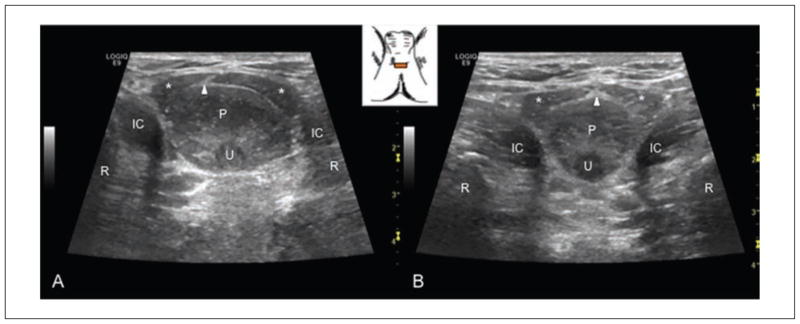
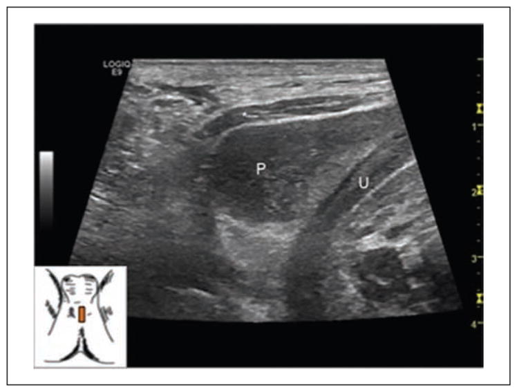
Idiopathic chronic male pelvic pain is difficult to diagnose and treat. Currently, diagnosis relies on subjective symptoms; objective measures of neuromuscular mechanisms have not been investigated. Sonographic imaging has been used to investigate these neuromuscular mechanisms in the female pelvic floor, but neither research nor books describe sonography evaluation of the male pelvic floor. The purpose of this study was to develop and evaluate a perineal sonographic technique for the examination of the male pelvic floor muscles. Anatomic landmarks were identified with images collected from two subjects, one with intermittent reports of pelvic pain and one with no history of pain in the pelvic region. A description of the equipment settings, the examination protocol, and the resulting comparative image analysis is included. A validated protocol such as this may be useful in documenting differences in the soft tissue structures between asymptomatic individuals and patients with chronic pelvic pain to aid in diagnosis and treatment. This is the first known study to report sonographic findings of the individual muscles in the male pelvic floor, and additional research is needed to validate the techniques that have been deemed feasible.
特发性慢性男性盆腔疼痛难以诊断和治疗。目前,诊断依赖于主观症状;尚未对神经肌肉机制的客观测量方法进行研究。超声成像已被用于研究女性盆底的这些神经肌肉机制,但无论是研究还是书籍都未描述男性盆底的超声检查评估。本研究的目的是开发并评估一种用于检查男性盆底肌肉的会阴超声技术。通过从两名受试者收集的图像确定了解剖标志,一名受试者间歇性报告盆腔疼痛,另一名受试者无盆腔疼痛病史。文中包括了设备设置、检查方案以及所得比较图像分析的描述。这样一种经过验证的方案可能有助于记录无症状个体与慢性盆腔疼痛患者之间软组织结构的差异,以辅助诊断和治疗。这是已知的第一项报告男性盆底各肌肉超声检查结果的研究,需要更多研究来验证已被认为可行的技术。