• 文献检索
  • 文档翻译
  • 深度研究
  • 学术资讯
  • Suppr Zotero 插件Zotero 插件
  • 邀请有礼
  • 套餐&价格
  • 历史记录
应用&插件
Suppr Zotero 插件Zotero 插件浏览器插件Mac 客户端Windows 客户端微信小程序
定价
高级版会员购买积分包购买API积分包
服务
文献检索文档翻译深度研究API 文档MCP 服务
关于我们
关于 Suppr公司介绍联系我们用户协议隐私条款
关注我们

Suppr 超能文献

核心技术专利:CN118964589B侵权必究
粤ICP备2023148730 号-1Suppr @ 2026

文献检索

告别复杂PubMed语法,用中文像聊天一样搜索,搜遍4000万医学文献。AI智能推荐,让科研检索更轻松。

立即免费搜索

文件翻译

保留排版,准确专业,支持PDF/Word/PPT等文件格式,支持 12+语言互译。

免费翻译文档

深度研究

AI帮你快速写综述,25分钟生成高质量综述,智能提取关键信息,辅助科研写作。

立即免费体验

基于亮度分布分析的脑淋巴瘤和胶质母细胞瘤分类。

Classification of cerebral lymphomas and glioblastomas featuring luminance distribution analysis.

机构信息

School of Electrical and Computer Engineering, Cornell University, Phillips Hall, Ithaca, NY 14853-5401, USA.

出版信息

Comput Math Methods Med. 2013;2013:619658. doi: 10.1155/2013/619658. Epub 2013 Jun 6.

DOI:10.1155/2013/619658
PMID:23840280
原文链接:https://pmc.ncbi.nlm.nih.gov/articles/PMC3690226/
Abstract

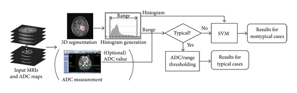
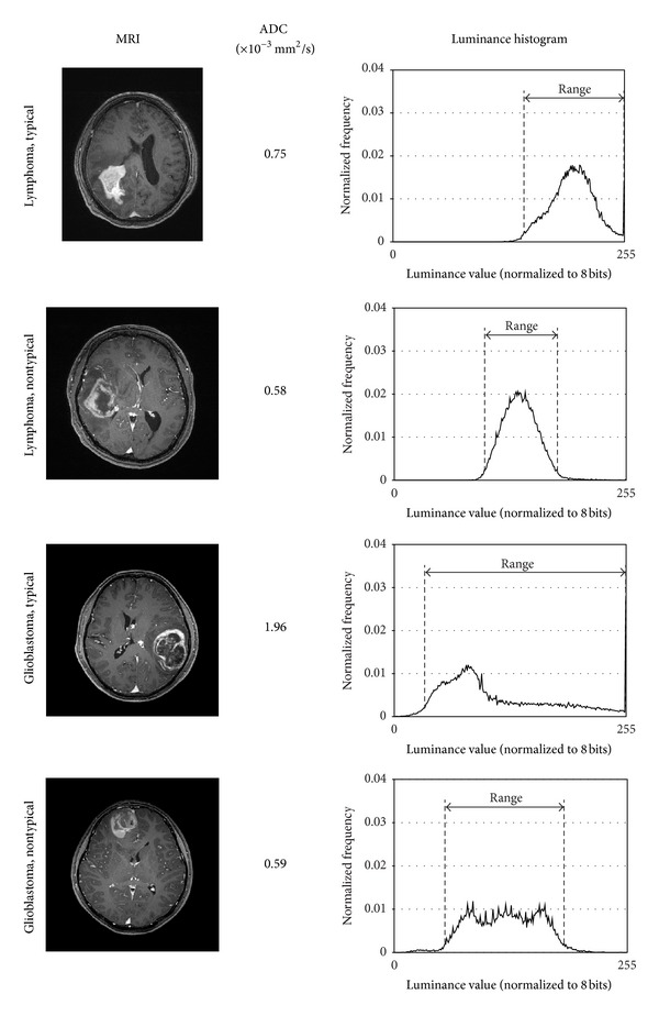
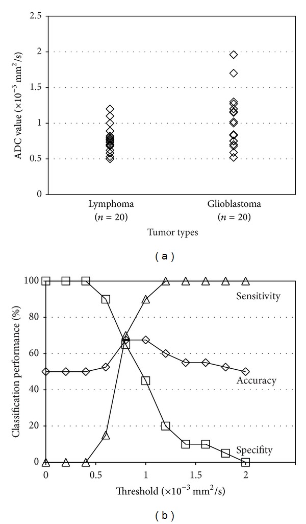
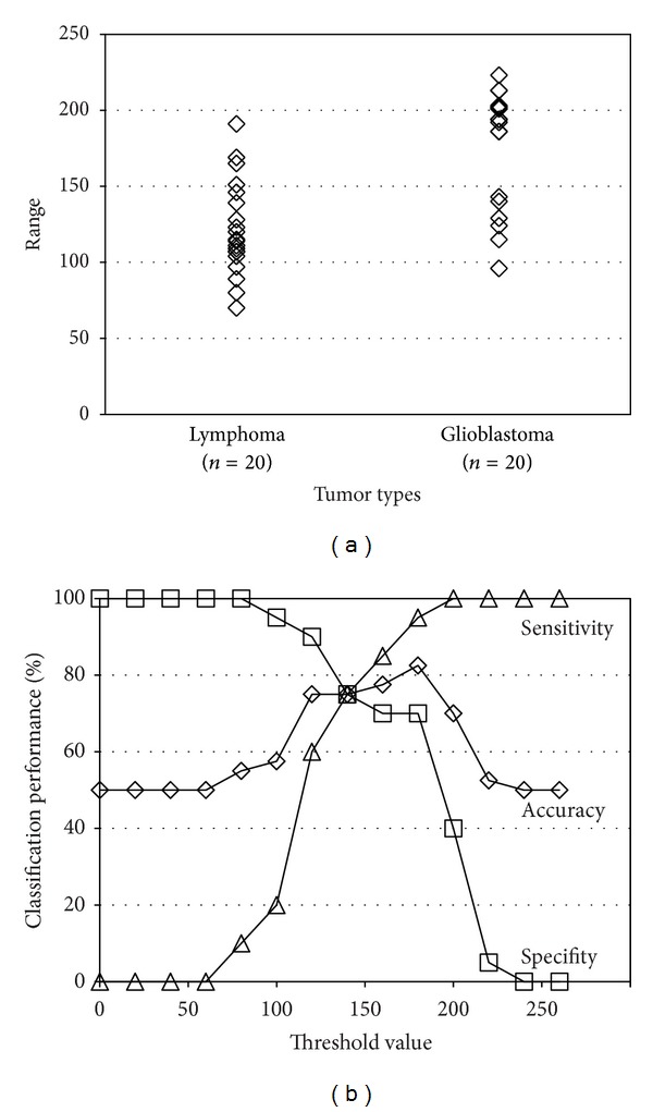
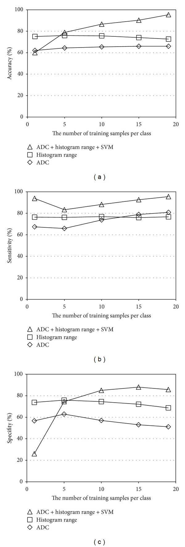
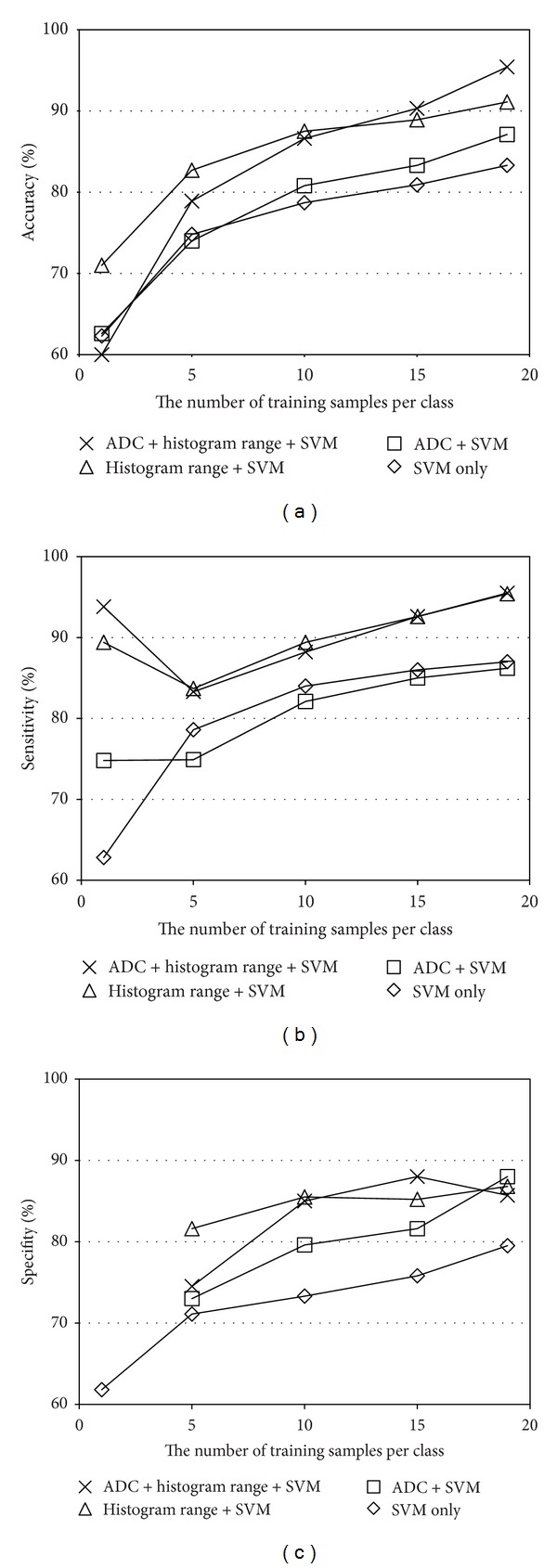
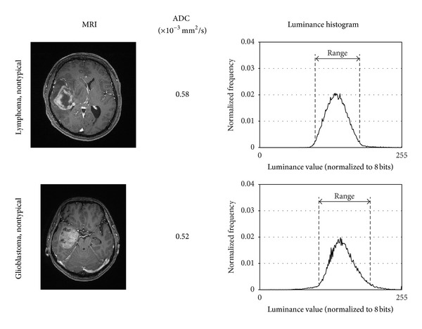
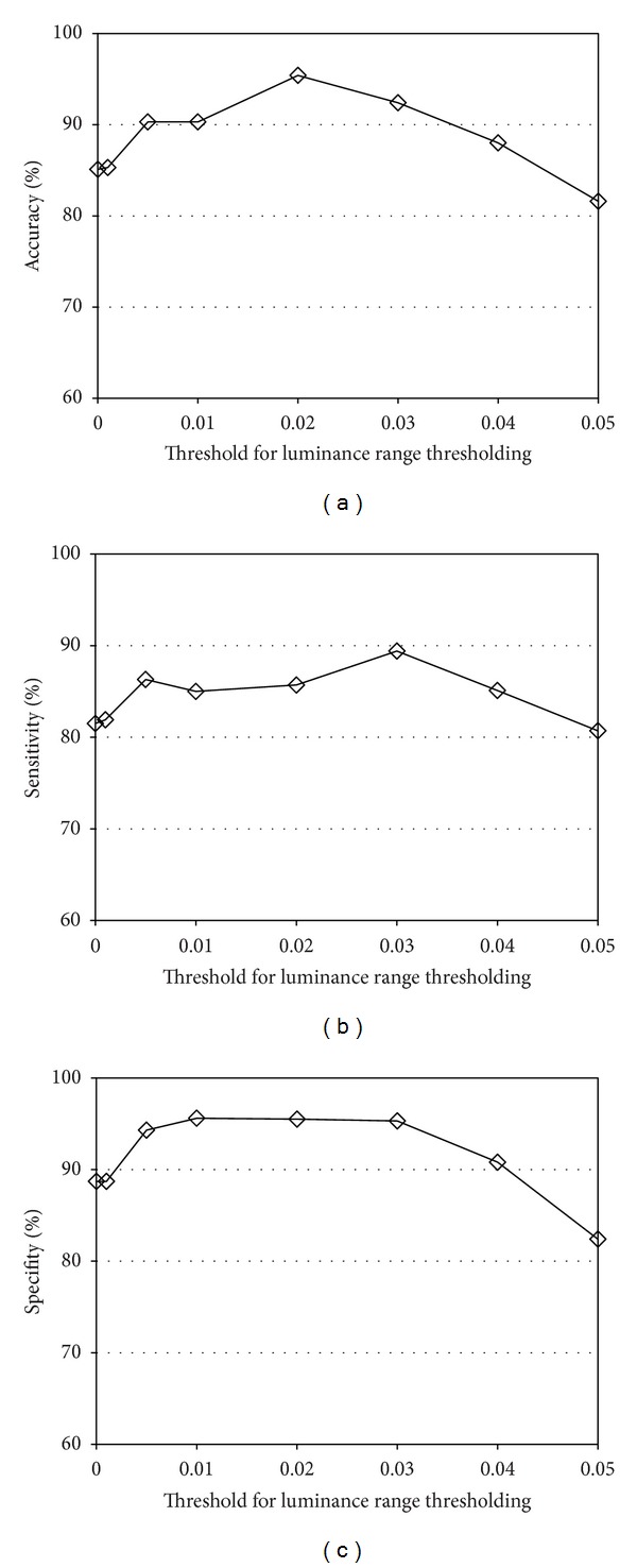
Differentiating lymphomas and glioblastomas is important for proper treatment planning. A number of works have been proposed but there are still some problems. For example, many works depend on thresholding a single feature value, which is susceptible to noise. In other cases, experienced observers are required to extract the feature values or to provide some interactions with the system. Even if experts are involved, interobserver variance becomes another problem. In addition, most of the works use only one or a few slice(s) because 3D tumor segmentation is time consuming. In this paper, we propose a tumor classification system that analyzes the luminance distribution of the whole tumor region. Typical cases are classified by the luminance range thresholding and the apparent diffusion coefficients (ADC) thresholding. Nontypical cases are classified by a support vector machine (SVM). Most of the processing elements are semiautomatic. Therefore, even novice users can use the system easily and get the same results as experts. The experiments were conducted using 40 MRI datasets. The classification accuracy of the proposed method was 91.1% without the ADC thresholding and 95.4% with the ADC thresholding. On the other hand, the baseline method, the conventional ADC thresholding, yielded only 67.5% accuracy.

摘要

区分淋巴瘤和胶质母细胞瘤对于制定正确的治疗计划非常重要。已经提出了许多方法,但仍然存在一些问题。例如,许多方法依赖于对单个特征值进行阈值处理,这容易受到噪声的影响。在其他情况下,需要有经验的观察者来提取特征值或与系统进行一些交互。即使有专家参与,观察者之间的差异也会成为另一个问题。此外,大多数方法只使用一个或几个切片,因为 3D 肿瘤分割非常耗时。在本文中,我们提出了一种肿瘤分类系统,该系统分析整个肿瘤区域的亮度分布。典型病例通过亮度范围阈值和表观扩散系数(ADC)阈值进行分类。非典型病例通过支持向量机(SVM)进行分类。大多数处理元素都是半自动的。因此,即使是新手用户也可以轻松使用该系统,并获得与专家相同的结果。实验使用了 40 个 MRI 数据集。所提出方法的分类准确率在没有 ADC 阈值的情况下为 91.1%,在有 ADC 阈值的情况下为 95.4%。另一方面,基线方法,即传统的 ADC 阈值处理,仅产生 67.5%的准确率。

https://cdn.ncbi.nlm.nih.gov/pmc/blobs/58ca/3690226/45951ca80d25/CMMM2013-619658.011.jpg
https://cdn.ncbi.nlm.nih.gov/pmc/blobs/58ca/3690226/88344f3482f6/CMMM2013-619658.001.jpg
https://cdn.ncbi.nlm.nih.gov/pmc/blobs/58ca/3690226/e2f67eb1554e/CMMM2013-619658.002.jpg
https://cdn.ncbi.nlm.nih.gov/pmc/blobs/58ca/3690226/564d5dbd0955/CMMM2013-619658.003.jpg
https://cdn.ncbi.nlm.nih.gov/pmc/blobs/58ca/3690226/4268d3cdf7d0/CMMM2013-619658.004.jpg
https://cdn.ncbi.nlm.nih.gov/pmc/blobs/58ca/3690226/25102cd9b244/CMMM2013-619658.005.jpg
https://cdn.ncbi.nlm.nih.gov/pmc/blobs/58ca/3690226/e13fc165dac4/CMMM2013-619658.006.jpg
https://cdn.ncbi.nlm.nih.gov/pmc/blobs/58ca/3690226/d83ec54cc475/CMMM2013-619658.007.jpg
https://cdn.ncbi.nlm.nih.gov/pmc/blobs/58ca/3690226/c24b0d6d4daf/CMMM2013-619658.008.jpg
https://cdn.ncbi.nlm.nih.gov/pmc/blobs/58ca/3690226/2adfd64827d7/CMMM2013-619658.009.jpg
https://cdn.ncbi.nlm.nih.gov/pmc/blobs/58ca/3690226/2dcc8399f3ad/CMMM2013-619658.010.jpg
https://cdn.ncbi.nlm.nih.gov/pmc/blobs/58ca/3690226/45951ca80d25/CMMM2013-619658.011.jpg

相似文献

1
Classification of cerebral lymphomas and glioblastomas featuring luminance distribution analysis.基于亮度分布分析的脑淋巴瘤和胶质母细胞瘤分类。
Comput Math Methods Med. 2013;2013:619658. doi: 10.1155/2013/619658. Epub 2013 Jun 6.
2
Accuracy of apparent diffusion coefficients and enhancement ratios on magnetic resonance imaging in differentiating primary cerebral lymphomas from glioblastoma.磁共振成像中表观扩散系数和增强率在鉴别原发性脑淋巴瘤与胶质母细胞瘤中的准确性
Neuroradiol J. 2019 Oct;32(5):328-334. doi: 10.1177/1971400919857556. Epub 2019 Jun 12.
3
Primary cerebral lymphoma and glioblastoma multiforme: differences in diffusion characteristics evaluated with diffusion tensor imaging.原发性脑淋巴瘤与多形性胶质母细胞瘤:通过扩散张量成像评估的扩散特征差异
AJNR Am J Neuroradiol. 2008 Mar;29(3):471-5. doi: 10.3174/ajnr.A0872. Epub 2007 Dec 7.
4
Differentiation of Glioblastoma and Lymphoma Using Feature Extraction and Support Vector Machine.使用特征提取和支持向量机对胶质母细胞瘤和淋巴瘤进行鉴别诊断。
CNS Neurol Disord Drug Targets. 2017;16(2):160-168. doi: 10.2174/1871527315666161018122909.
5
Perfusion and diffusion MR imaging in enhancing malignant cerebral tumors.增强型恶性脑肿瘤的灌注与扩散磁共振成像
Eur J Radiol. 2006 Jun;58(3):394-403. doi: 10.1016/j.ejrad.2005.12.032. Epub 2006 Mar 9.
6
Differentiation between Glioblastoma Multiforme and Primary Cerebral Lymphoma: Additional Benefits of Quantitative Diffusion-Weighted MR Imaging.多形性胶质母细胞瘤与原发性脑淋巴瘤的鉴别:定量扩散加权磁共振成像的额外优势
PLoS One. 2016 Sep 15;11(9):e0162565. doi: 10.1371/journal.pone.0162565. eCollection 2016.
7
Lymphomas and glioblastomas: differences in the apparent diffusion coefficient evaluated with high b-value diffusion-weighted magnetic resonance imaging at 3T.淋巴瘤和胶质母细胞瘤:3T 高 b 值扩散加权磁共振成像评估表观扩散系数的差异。
Eur J Radiol. 2012 Feb;81(2):339-44. doi: 10.1016/j.ejrad.2010.11.005. Epub 2010 Dec 3.
8
Diffusion radiomics as a diagnostic model for atypical manifestation of primary central nervous system lymphoma: development and multicenter external validation.扩散放射组学作为原发性中枢神经系统淋巴瘤不典型表现的诊断模型:开发和多中心外部验证。
Neuro Oncol. 2018 Aug 2;20(9):1251-1261. doi: 10.1093/neuonc/noy021.
9
Does adding FDG-PET to MRI improve the differentiation between primary cerebral lymphoma and glioblastoma? Observer performance study.FDG-PET 联合 MRI 是否能提高原发性脑淋巴瘤和胶质母细胞瘤的鉴别诊断能力?观察者性能研究。
Ann Nucl Med. 2011 Jul;25(6):432-8. doi: 10.1007/s12149-011-0483-1. Epub 2011 Mar 16.
10
Glioblastoma multiforme with atypical diffusion-weighted MR findings.具有非典型扩散加权磁共振成像表现的多形性胶质母细胞瘤
Br J Radiol. 2005 Nov;78(935):989-92. doi: 10.1259/bjr/12830378.

引用本文的文献

1
Artificial intelligence in the radiomic analysis of glioblastomas: A review, taxonomy, and perspective.胶质母细胞瘤放射组学分析中的人工智能:综述、分类及展望
Front Oncol. 2022 Aug 2;12:924245. doi: 10.3389/fonc.2022.924245. eCollection 2022.
2
Machine Learning in Differentiating Gliomas from Primary CNS Lymphomas: A Systematic Review, Reporting Quality, and Risk of Bias Assessment.机器学习在鉴别脑胶质瘤与原发性中枢神经系统淋巴瘤中的应用:系统综述、报告质量和偏倚风险评估。
AJNR Am J Neuroradiol. 2022 Apr;43(4):526-533. doi: 10.3174/ajnr.A7473. Epub 2022 Mar 31.
3
F-FDG-PET-based radiomics features to distinguish primary central nervous system lymphoma from glioblastoma.

本文引用的文献

1
Does adding FDG-PET to MRI improve the differentiation between primary cerebral lymphoma and glioblastoma? Observer performance study.FDG-PET 联合 MRI 是否能提高原发性脑淋巴瘤和胶质母细胞瘤的鉴别诊断能力?观察者性能研究。
Ann Nucl Med. 2011 Jul;25(6):432-8. doi: 10.1007/s12149-011-0483-1. Epub 2011 Mar 16.
2
Lymphomas and glioblastomas: differences in the apparent diffusion coefficient evaluated with high b-value diffusion-weighted magnetic resonance imaging at 3T.淋巴瘤和胶质母细胞瘤:3T 高 b 值扩散加权磁共振成像评估表观扩散系数的差异。
Eur J Radiol. 2012 Feb;81(2):339-44. doi: 10.1016/j.ejrad.2010.11.005. Epub 2010 Dec 3.
3
基于 F-FDG-PET 的影像组学特征可用于区分原发性中枢神经系统淋巴瘤和胶质母细胞瘤。
Neuroimage Clin. 2019;23:101912. doi: 10.1016/j.nicl.2019.101912. Epub 2019 Jun 27.
4
Accuracy of apparent diffusion coefficients and enhancement ratios on magnetic resonance imaging in differentiating primary cerebral lymphomas from glioblastoma.磁共振成像中表观扩散系数和增强率在鉴别原发性脑淋巴瘤与胶质母细胞瘤中的准确性
Neuroradiol J. 2019 Oct;32(5):328-334. doi: 10.1177/1971400919857556. Epub 2019 Jun 12.
5
Magnetic resonance texture analysis utility in differentiating intraparenchymal neurosarcoidosis from primary central nervous system lymphoma: a preliminary analysis.磁共振纹理分析在鉴别脑实质内结节病与原发性中枢神经系统淋巴瘤中的应用:一项初步分析
Neuroradiol J. 2019 Jun;32(3):203-209. doi: 10.1177/1971400919830173. Epub 2019 Feb 21.
6
Radiomics features to distinguish glioblastoma from primary central nervous system lymphoma on multi-parametric MRI.基于多参数MRI的区分胶质母细胞瘤与原发性中枢神经系统淋巴瘤的影像组学特征
Neuroradiology. 2018 Dec;60(12):1297-1305. doi: 10.1007/s00234-018-2091-4. Epub 2018 Sep 19.
7
Machine Learning-based Texture Analysis of Contrast-enhanced MR Imaging to Differentiate between Glioblastoma and Primary Central Nervous System Lymphoma.基于机器学习的对比增强磁共振成像纹理分析鉴别胶质母细胞瘤和原发性中枢神经系统淋巴瘤。
Magn Reson Med Sci. 2019 Jan 10;18(1):44-52. doi: 10.2463/mrms.mp.2017-0178. Epub 2018 May 16.
8
Primary central nervous system lymphoma and atypical glioblastoma: Differentiation using radiomics approach.原发性中枢神经系统淋巴瘤和非典型性脑胶质瘤:基于放射组学方法的鉴别诊断。
Eur Radiol. 2018 Sep;28(9):3832-3839. doi: 10.1007/s00330-018-5368-4. Epub 2018 Apr 6.
9
Differentiation of Enhancing Glioma and Primary Central Nervous System Lymphoma by Texture-Based Machine Learning.基于纹理的机器学习对强化型胶质瘤和原发性中枢神经系统淋巴瘤的鉴别
AJNR Am J Neuroradiol. 2017 Jun;38(6):1145-1150. doi: 10.3174/ajnr.A5173. Epub 2017 Apr 27.
Differentiation among glioblastoma multiforme, solitary metastatic tumor, and lymphoma using whole-tumor histogram analysis of the normalized cerebral blood volume in enhancing and perienhancing lesions.
使用增强和增强后病变的归一化脑血容量全瘤直方图分析鉴别多形性胶质母细胞瘤、单发转移性肿瘤和淋巴瘤。
AJNR Am J Neuroradiol. 2010 Oct;31(9):1699-706. doi: 10.3174/ajnr.A2161. Epub 2010 Jun 25.
4
Performance evaluation of radiologists with artificial neural network for differential diagnosis of intra-axial cerebral tumors on MR images.基于人工神经网络的放射科医生对磁共振成像上脑内肿瘤进行鉴别诊断的性能评估
AJNR Am J Neuroradiol. 2008 Jun;29(6):1153-8. doi: 10.3174/ajnr.A1037. Epub 2008 Apr 3.
5
Primary cerebral lymphoma and glioblastoma multiforme: differences in diffusion characteristics evaluated with diffusion tensor imaging.原发性脑淋巴瘤与多形性胶质母细胞瘤:通过扩散张量成像评估的扩散特征差异
AJNR Am J Neuroradiol. 2008 Mar;29(3):471-5. doi: 10.3174/ajnr.A0872. Epub 2007 Dec 7.
6
Perfusion and diffusion MR imaging in enhancing malignant cerebral tumors.增强型恶性脑肿瘤的灌注与扩散磁共振成像
Eur J Radiol. 2006 Jun;58(3):394-403. doi: 10.1016/j.ejrad.2005.12.032. Epub 2006 Mar 9.
7
Distinguishing of primary cerebral lymphoma from high-grade glioma with perfusion-weighted magnetic resonance imaging.利用灌注加权磁共振成像鉴别原发性脑淋巴瘤与高级别胶质瘤
Neurosci Lett. 2003 Feb 27;338(2):119-22. doi: 10.1016/s0304-3940(02)01367-8.