Department of Computer Science & Information Engineering, National Cheng Kung University, Tainan 701, Taiwan.
Comput Math Methods Med. 2013;2013:914124. doi: 10.1155/2013/914124. Epub 2013 Jun 6.
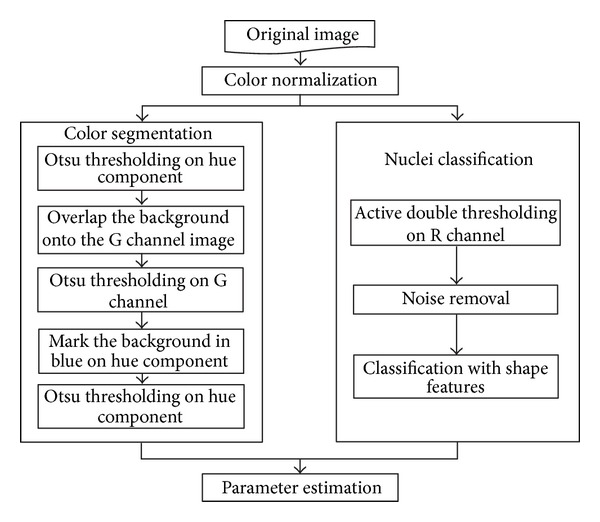
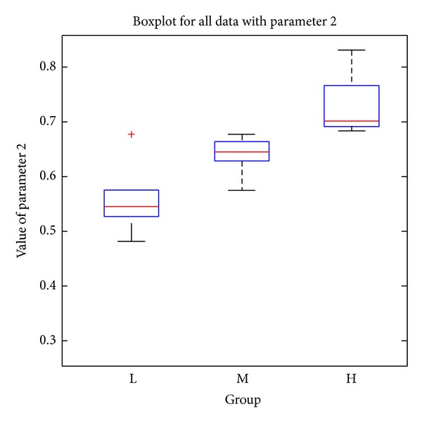
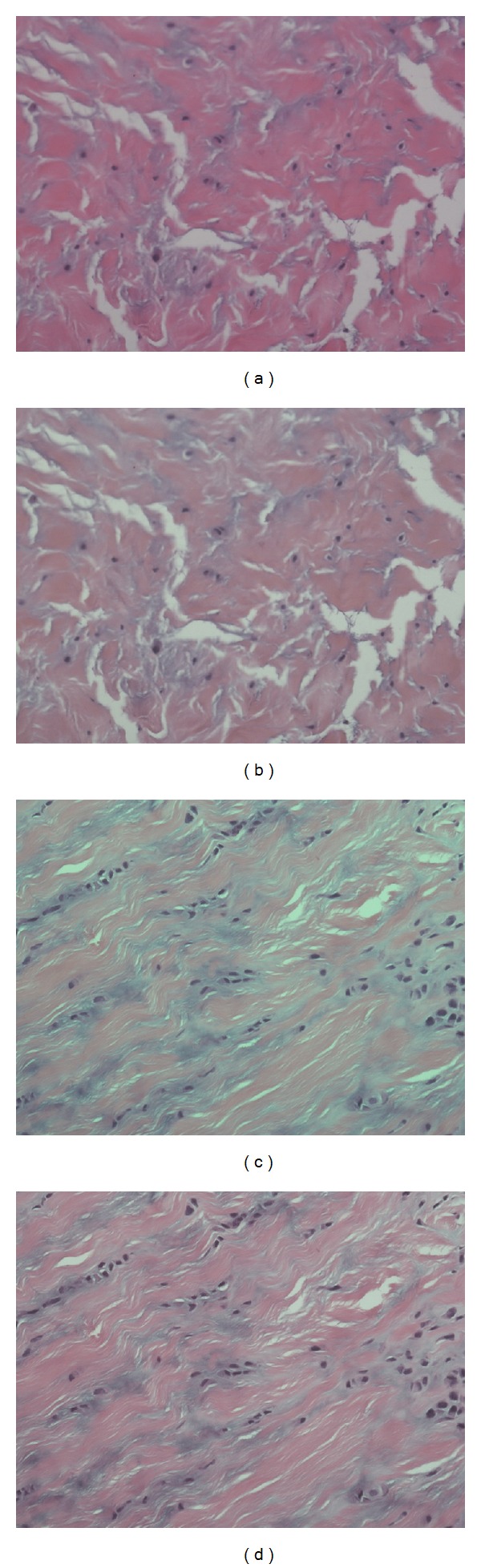
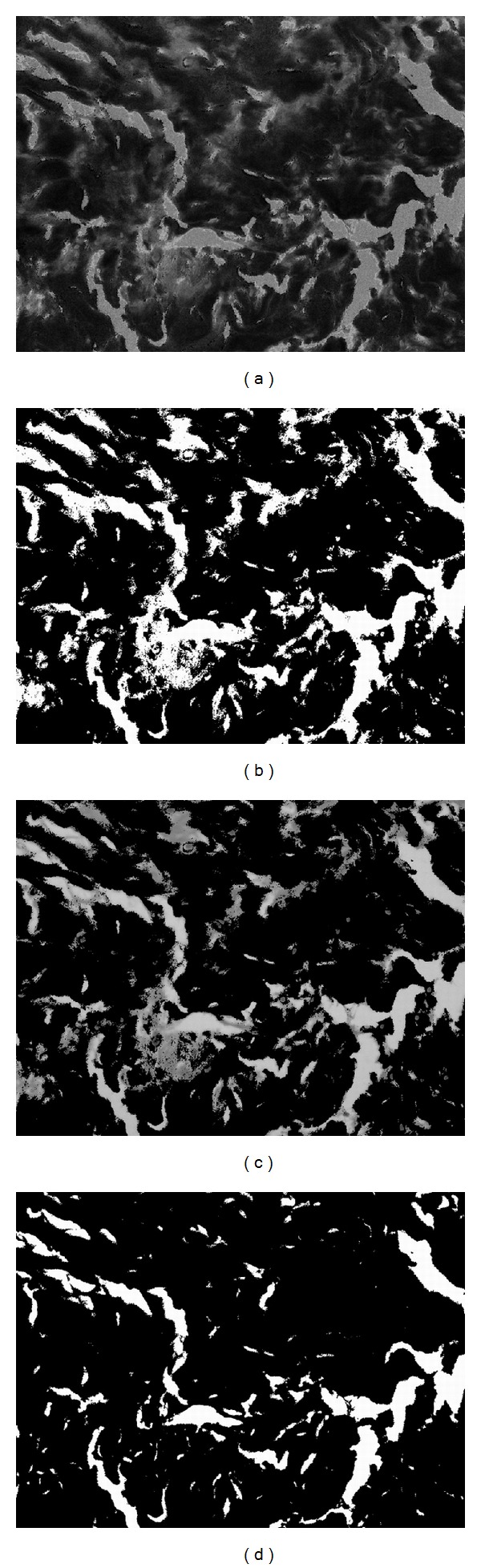
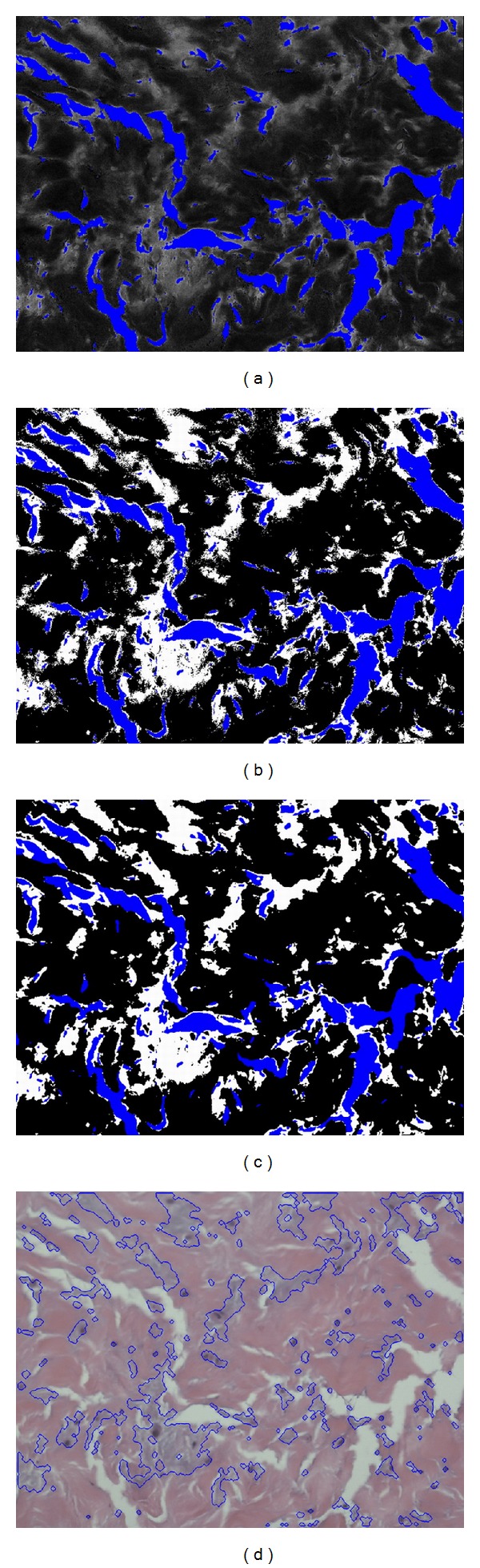
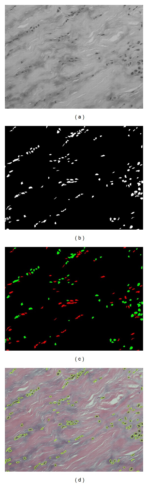
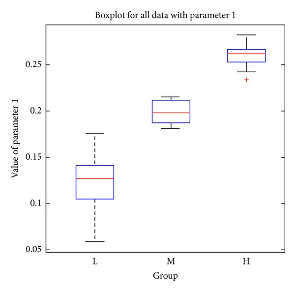
Quantifying the pathological features of flexor tendon pulleys is essential for grading the trigger finger since it provides clinicians with objective evidence derived from microscopic images. Although manual grading is time consuming and dependent on the observer experience, there is a lack of image processing methods for automatically extracting pulley pathological features. In this paper, we design and develop a color-based image segmentation system to extract the color and shape features from pulley microscopic images. Two parameters which are the size ratio of abnormal tissue regions and the number ratio of abnormal nuclei are estimated as the pathological progression indices. The automatic quantification results show clear discrimination among different levels of diseased pulley specimens which are prone to misjudgments for human visual inspection. The proposed system provides a reliable and automatic way to obtain pathological parameters instead of manual evaluation which is with intra- and interoperator variability. Experiments with 290 microscopic images from 29 pulley specimens show good correspondence with pathologist expectations. Hence, the proposed system has great potential for assisting clinical experts in routine histopathological examinations.
量化屈肌腱滑车的病理特征对于扳机指的分级至关重要,因为它为临床医生提供了源自显微镜图像的客观证据。虽然手动分级耗时且依赖于观察者的经验,但缺乏用于自动提取滑车病理特征的图像处理方法。在本文中,我们设计并开发了一种基于颜色的图像分割系统,以从滑车显微镜图像中提取颜色和形状特征。估计两个参数,即异常组织区域的大小比和异常核的数量比,作为病理进展指标。自动量化结果显示,不同病变程度的滑车标本之间有明显的区分,而人工视觉检查容易出现误判。与手动评估相比,所提出的系统提供了一种可靠且自动的获取病理参数的方法,具有内在和操作者间的可变性。对 29 个滑车标本的 290 张显微镜图像进行的实验表明,与病理学家的预期有很好的一致性。因此,该系统有望辅助临床专家进行常规组织病理学检查。