UCL Institute of Ophthalmology, Ocular Biology and Therapeutics, London, United Kingdom.
PLoS One. 2013 Jun 28;8(6):e67263. doi: 10.1371/journal.pone.0067263. Print 2013.
With ageing extracellular material is deposited in Bruch's membrane, as drusen. Lipofuscin is deposited in retinal pigment epithelial cells. Both of these changes are associated with age related macular degeneration, a disease now believed to involve chronic inflammation at the retinal-choroidal interface. We hypothesise that these molecules may act as danger signals, causing the production of inflammatory chemokines and cytokines by the retinal pigment epithelium, via activation of pattern recognition receptors.
ARPE-19 cells were stimulated in vitro with the following reported components of drusen: amyloid-ß (1-42), Carboxyethylpyrrole (CEP) modified proteins (CEP-HSA), Nε-(Carboxymethyl)lysine (CML) modified proteins and aggregated vitronectin. The cells were also stimulated with the major fluorophore of lipofuscin: N-retinylidene-N-retinylethanolamine (A2E). Inflammatory chemokine and cytokine production was assessed using Multiplex assays and ELISA. The mechanistic evaluation of the NLRP3 inflammasome pathway was assessed in a stepwise fashion.
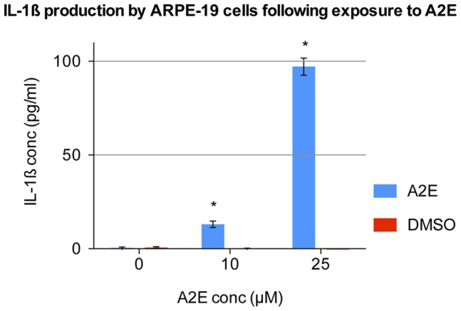
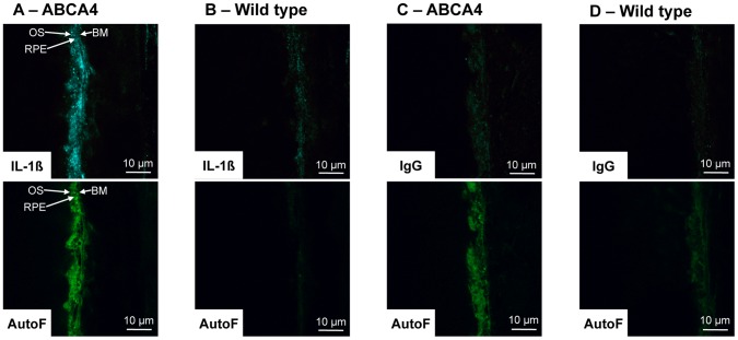
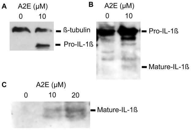
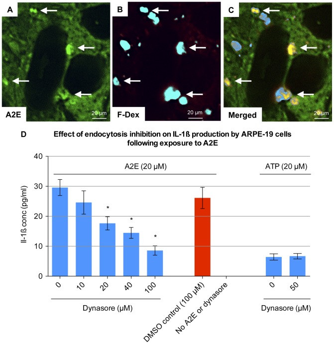
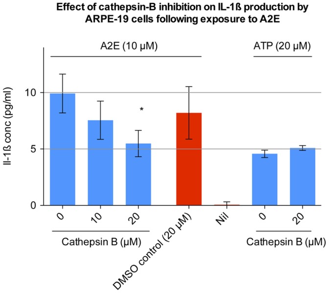
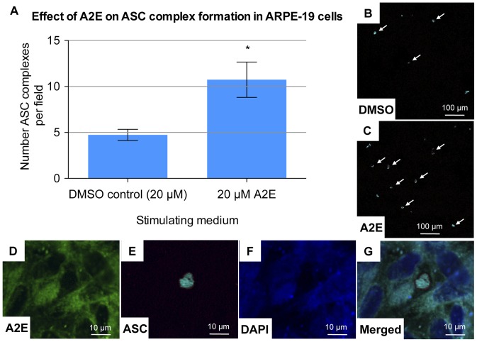
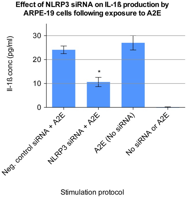
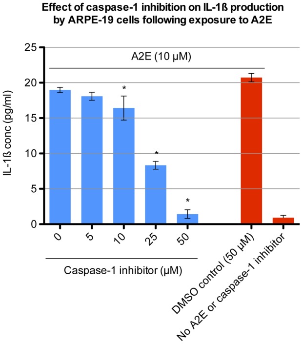
Of all the molecules tested only A2E induced inflammatory chemokine and cytokine production. 25 µM A2E induced the production of significantly increased levels of the chemokines IL-8, MCP-1, MCG and MIP-1α, the cytokines IL-1ß, IL-2, IL-6, and TNF-α, and the protein VEGF-A. The release of IL-1ß was studied further, and was determined to be due to NLRP3 inflammasome activation. The pathway of activation involved endocytosis of A2E, and the three inflammasome components NLRP3, ASC and activated caspase-1. Immunohistochemical staining of ABCA4 knockout mice, which show progressive accumulation of A2E levels with age, showed increased amounts of IL-1ß proximal to the retinal pigment epithelium.
A2E has the ability to stimulate inflammatory chemokine and cytokine production by RPE cells. The pattern recognition receptor NLRP3 is involved in this process. This provides further evidence for the link between A2E, inflammation, and the pathogenesis of AMD. It also supports the recent discovery of NLRP3 inflammasome activation in AMD.
随着细胞外物质在布鲁赫膜中沉积(如玻璃膜疣)和脂褐素在视网膜色素上皮细胞中的沉积,与年龄相关的黄斑变性(AMD)相关。现在认为这种疾病涉及视网膜脉络膜界面的慢性炎症。我们假设这些分子可能作为危险信号,通过激活模式识别受体,导致视网膜色素上皮细胞产生炎症趋化因子和细胞因子。
体外用以下报道的玻璃膜疣成分刺激 ARPE-19 细胞:淀粉样蛋白-β(1-42)、羧乙基吡咯(CEP)修饰蛋白(CEP-HSA)、Nε-(羧甲基)赖氨酸(CML)修饰蛋白和聚集的玻连蛋白。还用脂褐素的主要荧光团:N-视黄基-N-视黄醇乙胺(A2E)刺激细胞。使用多重分析和 ELISA 评估炎症趋化因子和细胞因子的产生。逐步评估 NLRP3 炎性体途径的机制评估。
在所测试的所有分子中,只有 A2E 诱导了炎症趋化因子和细胞因子的产生。25μMA2E 诱导产生显著增加的趋化因子 IL-8、MCP-1、MCG 和 MIP-1α、细胞因子 IL-1β、IL-2、IL-6 和 TNF-α以及蛋白 VEGF-A。进一步研究了 IL-1β的释放,发现这是由于 NLRP3 炎性体的激活。激活途径涉及 A2E 的内吞作用,以及 NLRP3、ASC 和活化的 caspase-1 三个炎性体成分。用 ABCA4 敲除小鼠进行免疫组织化学染色,该小鼠随着年龄的增长逐渐积累 A2E 水平,结果显示在视网膜色素上皮附近有更多的 IL-1β。
A2E 具有刺激 RPE 细胞产生炎症趋化因子和细胞因子的能力。模式识别受体 NLRP3 参与了这一过程。这为 A2E、炎症和 AMD 发病机制之间的联系提供了进一步的证据。它还支持最近在 AMD 中发现 NLRP3 炎性体的激活。