• 文献检索
  • 文档翻译
  • 深度研究
  • 学术资讯
  • Suppr Zotero 插件Zotero 插件
  • 邀请有礼
  • 套餐&价格
  • 历史记录
应用&插件
Suppr Zotero 插件Zotero 插件浏览器插件Mac 客户端Windows 客户端微信小程序
定价
高级版会员购买积分包购买API积分包
服务
文献检索文档翻译深度研究API 文档MCP 服务
关于我们
关于 Suppr公司介绍联系我们用户协议隐私条款
关注我们

Suppr 超能文献

核心技术专利:CN118964589B侵权必究
粤ICP备2023148730 号-1Suppr @ 2026

文献检索

告别复杂PubMed语法,用中文像聊天一样搜索,搜遍4000万医学文献。AI智能推荐,让科研检索更轻松。

立即免费搜索

文件翻译

保留排版,准确专业,支持PDF/Word/PPT等文件格式,支持 12+语言互译。

免费翻译文档

深度研究

AI帮你快速写综述,25分钟生成高质量综述,智能提取关键信息,辅助科研写作。

立即免费体验

双回波动态对比增强 MRI 鉴别乳腺癌与纤维腺瘤。

Differentiation of breast cancer from fibroadenoma with dual-echo dynamic contrast-enhanced MRI.

机构信息

Department of Radiology, First Affiliated Hospital of Zhejiang Chinese Medical University, Hangzhou, Zhejiang, China.

出版信息

PLoS One. 2013 Jul 2;8(7):e67731. doi: 10.1371/journal.pone.0067731. Print 2013.

DOI:10.1371/journal.pone.0067731
PMID:23844077
原文链接:https://pmc.ncbi.nlm.nih.gov/articles/PMC3699626/
Abstract

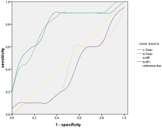
Dynamic contrast-enhanced magnetic resonance imaging (DCE MRI) of the breast is a routinely used imaging method which is highly sensitive for detecting breast malignancy. Specificity, though, remains suboptimal. Dynamic susceptibility contrast magnetic resonance imaging (DSC MRI), an alternative dynamic contrast imaging technique, evaluates perfusion-related parameters unique from DCE MRI. Previous work has shown that the combination of DSC MRI with DCE MRI can improve diagnostic specificity, though an additional administration of intravenous contrast is required. Dual-echo MRI can measure both T1W DCE MRI and T2W DSC MRI parameters with a single contrast bolus, but has not been previously implemented in breast imaging. We have developed a dual-echo gradient-echo sequence to perform such simultaneous measurements in the breast, and use it to calculate the semi-quantitative T1W and T2W related parameters such as peak enhancement ratio, time of maximal enhancement, regional blood flow, and regional blood volume in 20 malignant lesions and 10 benign fibroadenomas in 38 patients. Imaging parameters were compared to surgical or biopsy obtained tissue samples. Receiver operating characteristic (ROC) curves and area under the ROC curves were calculated for each parameter and combination of parameters. The time of maximal enhancement derived from DCE MRI had a 90% sensitivity and 69% specificity for predicting malignancy. When combined with DSC MRI derived regional blood flow and volume parameters, sensitivity remained unchanged at 90% but specificity increased to 80%. In conclusion, we show that dual-echo MRI with a single administration of contrast agent can simultaneously measure both T1W and T2*W related perfusion and kinetic parameters in the breast and the combination of DCE MRI and DSC MRI parameters improves the diagnostic performance of breast MRI to differentiate breast cancer from benign fibroadenomas.

摘要

动态对比增强磁共振成像(DCE MRI)是一种常规使用的成像方法,对检测乳腺癌具有高度敏感性。然而,其特异性仍然不理想。动态磁化率对比磁共振成像(DSC MRI)是一种替代的动态对比成像技术,可评估与灌注相关的、与 DCE MRI 不同的参数。先前的工作表明,DSC MRI 与 DCE MRI 的结合可以提高诊断的特异性,尽管需要额外的静脉内造影剂注射。双回波磁共振成像可以用单次造影剂注射测量 T1W DCE MRI 和 T2W DSC MRI 参数,但以前并未在乳腺成像中实施。我们开发了一种双回波梯度回波序列,可以在乳腺中进行这种同时测量,并使用它来计算半定量 T1W 和 T2W 相关参数,如峰值增强比、最大增强时间、区域血流量和 38 例患者的 20 例恶性病变和 10 例良性纤维腺瘤中的区域血容量。对成像参数与手术或活检获得的组织样本进行了比较。为每个参数和参数组合计算了接收器工作特性(ROC)曲线和 ROC 曲线下面积。从 DCE MRI 得出的最大增强时间对预测恶性肿瘤具有 90%的敏感性和 69%的特异性。当与 DSC MRI 得出的区域血流和容积参数结合时,敏感性保持不变,为 90%,但特异性提高到 80%。总之,我们表明,单次造影剂注射的双回波 MRI 可以同时测量乳腺中的 T1W 和 T2*W 相关灌注和动力学参数,DCE MRI 和 DSC MRI 参数的组合提高了乳腺 MRI 鉴别乳腺癌与良性纤维腺瘤的诊断性能。

https://cdn.ncbi.nlm.nih.gov/pmc/blobs/9c13/3699626/fe42676e916c/pone.0067731.g001.jpg

相似文献

1
Differentiation of breast cancer from fibroadenoma with dual-echo dynamic contrast-enhanced MRI.双回波动态对比增强 MRI 鉴别乳腺癌与纤维腺瘤。
PLoS One. 2013 Jul 2;8(7):e67731. doi: 10.1371/journal.pone.0067731. Print 2013.
2
Breast lesions: evaluation with dynamic contrast-enhanced T1-weighted MR imaging and with T2*-weighted first-pass perfusion MR imaging.乳腺病变:采用动态对比增强T1加权磁共振成像及T2*加权首过灌注磁共振成像进行评估
Radiology. 2000 Aug;216(2):545-53. doi: 10.1148/radiology.216.2.r00au36545.
3
Intravoxel incoherent motion MR imaging for breast lesions: comparison and correlation with pharmacokinetic evaluation from dynamic contrast-enhanced MR imaging.体素内不相干运动磁共振成像用于乳腺病变:与动态对比增强磁共振成像的药代动力学评估的比较及相关性
Eur Radiol. 2016 Nov;26(11):3888-3898. doi: 10.1007/s00330-016-4241-6. Epub 2016 Feb 10.
4
Automatic identification and classification of characteristic kinetic curves of breast lesions on DCE-MRI.动态对比增强磁共振成像(DCE-MRI)中乳腺病变特征性动力学曲线的自动识别与分类
Med Phys. 2006 Aug;33(8):2878-87. doi: 10.1118/1.2210568.
5
Differentiating benign and malignant breast lesions with T2*-weighted first pass perfusion imaging.利用T2*加权首过灌注成像鉴别乳腺良恶性病变
Acta Radiol. 1999 Jan;40(1):45-51. doi: 10.1080/02841859909174402.
6
Optimization of time-to-peak analysis for differentiating malignant and benign breast lesions with dynamic contrast-enhanced MRI.优化时间至峰值分析,以区分动态对比增强 MRI 中的良恶性乳腺病变。
Acad Radiol. 2011 Jun;18(6):694-704. doi: 10.1016/j.acra.2011.01.005. Epub 2011 Mar 21.
7
Dynamic MR imaging of the breast combined with analysis of contrast agent kinetics in the differentiation of primary breast tumours.
Clin Radiol. 1997 Jul;52(7):516-26. doi: 10.1016/s0009-9260(97)80328-5.
8
Differentiation between malignant and benign breast masses: combination of semi-quantitative analysis on DCE-MRI and histogram analysis of ADC maps.乳腺良恶性肿块的鉴别:动态对比增强磁共振成像(DCE-MRI)的半定量分析与表观扩散系数(ADC)图的直方图分析相结合
Clin Radiol. 2018 May;73(5):460-466. doi: 10.1016/j.crad.2017.11.026. Epub 2017 Dec 30.
9
Characterization of spatiotemporal changes for the classification of dynamic contrast-enhanced magnetic-resonance breast lesions.动态对比增强磁共振乳腺病变分类的时空变化特征。
Artif Intell Med. 2013 Jun;58(2):101-14. doi: 10.1016/j.artmed.2013.03.002. Epub 2013 Mar 30.
10
Breast DCE-MRI: influence of postcontrast timing on automated lesion kinetics assessments and discrimination of benign and malignant lesions.乳腺动态对比增强磁共振成像:对比剂注射后时间对病变自动动力学评估及良恶性病变鉴别的影响
Acad Radiol. 2014 Sep;21(9):1195-203. doi: 10.1016/j.acra.2014.04.013. Epub 2014 Jul 4.

引用本文的文献

1
Development of a Non-invasive Assessment of Hypoxia and Neovascularization with Magnetic Resonance Imaging in Benign and Malignant Breast Tumors: Initial Results.良性和恶性乳腺肿瘤的磁共振成像非侵入性缺氧和新生血管评估的发展:初步结果。
Mol Imaging Biol. 2019 Aug;21(4):758-770. doi: 10.1007/s11307-018-1298-4.
2
Diagnostic value of dynamic contrast-enhanced magnetic resonance imaging in rectal cancer and its correlation with tumor differentiation.动态对比增强磁共振成像在直肠癌中的诊断价值及其与肿瘤分化的相关性
Mol Clin Oncol. 2016 Apr;4(4):500-506. doi: 10.3892/mco.2016.762. Epub 2016 Feb 3.

本文引用的文献

1
Quantifying tumor vascular heterogeneity with dynamic contrast-enhanced magnetic resonance imaging: a review.利用动态对比增强磁共振成像量化肿瘤血管异质性:综述
J Biomed Biotechnol. 2011;2011:732848. doi: 10.1155/2011/732848. Epub 2011 Apr 26.
2
Feasibility of high temporal resolution breast DCE-MRI using compressed sensing theory.应用压缩感知理论实现高时间分辨率的乳腺 DCE-MRI 可行性研究。
Med Phys. 2010 Sep;37(9):4971-81. doi: 10.1118/1.3483094.
3
Quantitative analysis of clinical dynamic contrast-enhanced MR imaging for evaluating treatment response in human breast cancer.
临床动态对比增强磁共振成像定量分析评估人乳腺癌治疗反应。
Radiology. 2010 Oct;257(1):47-55. doi: 10.1148/radiol.10092169. Epub 2010 Aug 16.
4
Is breast MRI helpful in the evaluation of inconclusive mammographic findings?乳腺磁共振成像(MRI)对评估乳腺钼靶检查结果不明确的情况有帮助吗?
AJR Am J Roentgenol. 2009 Oct;193(4):986-93. doi: 10.2214/AJR.08.1229.
5
A clinically feasible method to estimate pharmacokinetic parameters in breast cancer.一种在乳腺癌中估算药代动力学参数的临床可行方法。
Med Phys. 2009 Aug;36(8):3786-94. doi: 10.1118/1.3152113.
6
Simultaneous quantification of perfusion and permeability in the prostate using dynamic contrast-enhanced magnetic resonance imaging with an inversion-prepared dual-contrast sequence.使用反转准备双对比序列的动态对比增强磁共振成像同时定量评估前列腺的灌注和通透性。
Ann Biomed Eng. 2009 Apr;37(4):749-62. doi: 10.1007/s10439-009-9645-x. Epub 2009 Jan 24.
7
Characterization of breast lesions with CE-MR multimodal morphological and kinetic analysis: comparison with conventional mammography and high-resolution ultrasound.对比传统乳腺钼靶摄影和高分辨率超声,采用CE-MR多模态形态学和动力学分析对乳腺病变进行特征描述
Eur J Radiol. 2009 Apr;70(1):69-76. doi: 10.1016/j.ejrad.2008.01.012. Epub 2008 Mar 4.
8
Meta-analysis of MR imaging in the diagnosis of breast lesions.磁共振成像在乳腺病变诊断中的Meta分析。
Radiology. 2008 Jan;246(1):116-24. doi: 10.1148/radiol.2461061298. Epub 2007 Nov 16.
9
Current status of breast MR imaging. Part 2. Clinical applications.乳腺磁共振成像的现状。第2部分。临床应用。
Radiology. 2007 Sep;244(3):672-91. doi: 10.1148/radiol.2443051661.
10
Quantification of perfusion and permeability in breast tumors with a deconvolution-based analysis of second-bolus T1-DCE data.基于去卷积的二次团注T1加权动态对比增强数据分析法对乳腺肿瘤灌注和通透性的定量分析
J Magn Reson Imaging. 2007 Jun;25(6):1159-67. doi: 10.1002/jmri.20937.