Department of Nephrology and Rheumatology, Georg-August University, Göttingen, Germany.
Department of Zoology, Faculty of Science, Alexandria University, Alexandria, Egypt.
PLoS One. 2013 Jul 11;8(7):e68301. doi: 10.1371/journal.pone.0068301. Print 2013.
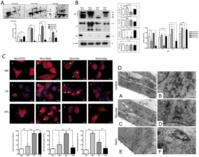
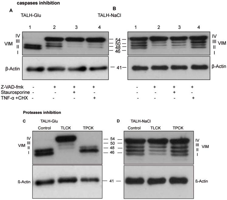
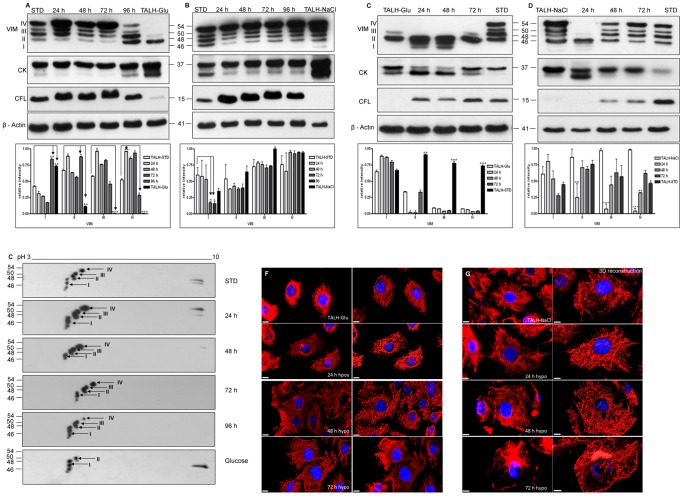
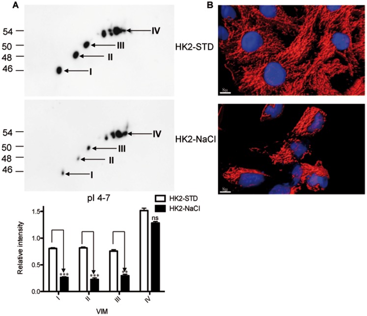
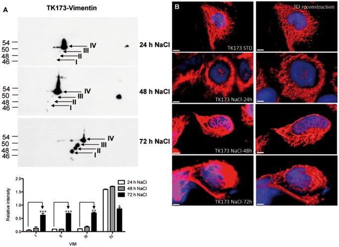
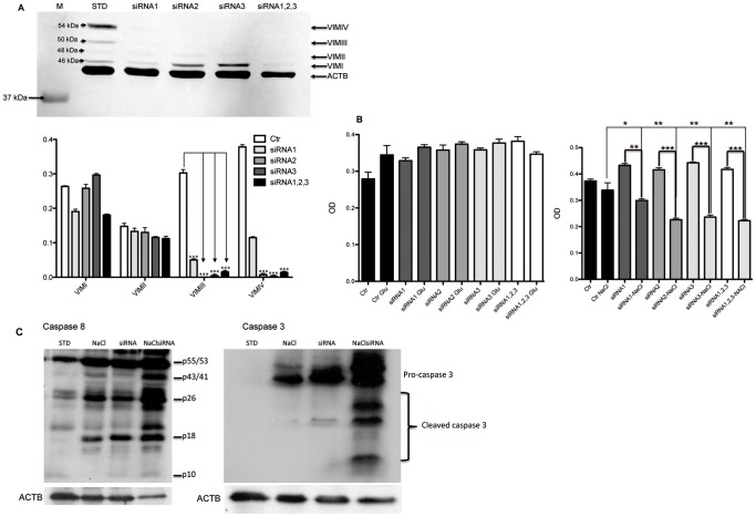
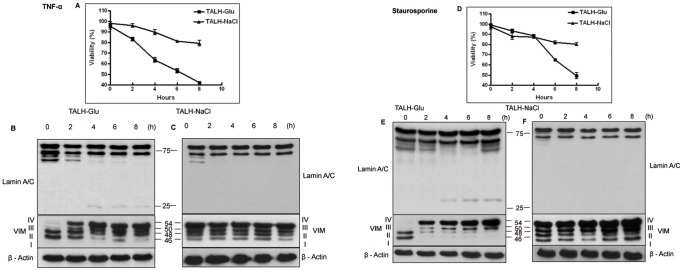
Osmotic stress has been shown to regulate cytoskeletal protein expression. It is generally known that vimentin is rapidly degraded during apoptosis by multiple caspases, resulting in diverse vimentin fragments. Despite the existence of the known apoptotic vimentin fragments, we demonstrated in our study the existence of different forms of vimentin VIM I, II, III, and IV with different molecular weights in various renal cell lines. Using a proteomics approach followed by western blot analyses and immunofluorescence staining, we proved the apoptosis-independent existence and differential regulation of different vimentin forms under varying conditions of osmolarity in renal cells. Similar impacts of osmotic stress were also observed on the expression of other cytoskeleton intermediate filament proteins; e.g., cytokeratin. Interestingly, 2D western blot analysis revealed that the forms of vimentin are regulated independently of each other under glucose and NaCl osmotic stress. Renal cells, adapted to high NaCl osmotic stress, express a high level of VIM IV (the form with the highest molecular weight), besides the three other forms, and exhibit higher resistance to apoptotic induction with TNF-α or staurosporin compared to the control. In contrast, renal cells that are adapted to high glucose concentration and express only the lower-molecular-weight forms VIM I and II, were more susceptible to apoptosis. Our data proved the existence of different vimentin forms, which play an important role in cell resistance to osmotic stress and are involved in cell protection against apoptosis.
渗透胁迫已被证明可调节细胞骨架蛋白的表达。众所周知,细胞凋亡过程中,多种半胱天冬酶迅速降解波形蛋白,导致出现多种不同的波形蛋白片段。尽管存在已知的凋亡性波形蛋白片段,但我们在本研究中证实,在不同的肾细胞系中存在不同形式的分子量不同的波形蛋白 VIM I、II、III 和 IV。通过采用蛋白质组学方法,结合 Western blot 分析和免疫荧光染色,我们证明了在渗透压条件不同的情况下,波形蛋白的存在形式具有凋亡独立性和差异调节性。其他细胞骨架中间丝蛋白如细胞角蛋白的表达也受到渗透胁迫的类似影响。有趣的是,2D Western blot 分析显示,在葡萄糖和 NaCl 渗透胁迫下,波形蛋白的形式彼此独立地受到调节。适应高 NaCl 渗透胁迫的肾细胞表达高水平的 VIM IV(分子量最高的形式),除了其他三种形式外,与对照相比,它们对 TNF-α或星形孢菌素诱导的凋亡具有更高的抗性。相比之下,适应高葡萄糖浓度且仅表达低分子量形式 VIM I 和 II 的肾细胞对凋亡更敏感。我们的数据证明了不同波形蛋白形式的存在,这些形式在细胞对渗透胁迫的抗性中发挥重要作用,并参与细胞对凋亡的保护。