• 文献检索
  • 文档翻译
  • 深度研究
  • 学术资讯
  • Suppr Zotero 插件Zotero 插件
  • 邀请有礼
  • 套餐&价格
  • 历史记录
应用&插件
Suppr Zotero 插件Zotero 插件浏览器插件Mac 客户端Windows 客户端微信小程序
定价
高级版会员购买积分包购买API积分包
服务
文献检索文档翻译深度研究API 文档MCP 服务
关于我们
关于 Suppr公司介绍联系我们用户协议隐私条款
关注我们

Suppr 超能文献

核心技术专利:CN118964589B侵权必究
粤ICP备2023148730 号-1Suppr @ 2026

文献检索

告别复杂PubMed语法,用中文像聊天一样搜索,搜遍4000万医学文献。AI智能推荐,让科研检索更轻松。

立即免费搜索

文件翻译

保留排版,准确专业,支持PDF/Word/PPT等文件格式,支持 12+语言互译。

免费翻译文档

深度研究

AI帮你快速写综述,25分钟生成高质量综述,智能提取关键信息,辅助科研写作。

立即免费体验

早期脑损伤:蛛网膜下腔出血研究的不断发展前沿。

Early brain injury, an evolving frontier in subarachnoid hemorrhage research.

机构信息

Departments of Neurosurgery, Physiology, and Anesthesiology, Loma Linda University, School of Medicine, Loma Linda, CA 92354, USA.

出版信息

Transl Stroke Res. 2013 Aug;4(4):432-46. doi: 10.1007/s12975-013-0257-2.

DOI:10.1007/s12975-013-0257-2
PMID:23894255
原文链接:https://pmc.ncbi.nlm.nih.gov/articles/PMC3719879/
Abstract

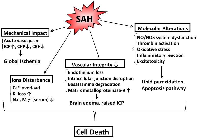
Subarachnoid hemorrhage (SAH), predominantly caused by a ruptured aneurysm, is a devastating neurological disease that has a morbidity and mortality rate higher than 50%. Most of the traditional in vivo research has focused on the pathophysiological or morphological changes of large-arteries after intracisternal blood injection. This was due to a widely held assumption that delayed vasospasm following SAH was the major cause of delayed cerebral ischemia and poor outcome. However, the results of the CONSCIOUS-1 trial implicated some other pathophysiological factors, independent of angiographic vasospasm, in contributing to the poor clinical outcome. The term early brain injury (EBI) has been coined and describes the immediate injury to the brain after SAH, before onset of delayed vasospasm. During the EBI period, a ruptured aneurysm brings on many physiological derangements such as increasing intracranial pressure (ICP), decreased cerebral blood flow (CBF), and global cerebral ischemia. These events initiate secondary injuries such as blood-brain barrier disruption, inflammation, and oxidative cascades that all ultimately lead to cell death. Given the fact that the reversal of vasospasm does not appear to improve patient outcome, it could be argued that the treatment of EBI may successfully attenuate some of the devastating secondary injuries and improve the outcome of patients with SAH. In this review, we provide an overview of the major advances in EBI after SAH research.

摘要

蛛网膜下腔出血(SAH)主要由破裂的动脉瘤引起,是一种毁灭性的神经系统疾病,其发病率和死亡率高于 50%。大多数传统的体内研究都集中在脑室内注血后大动脉的病理生理或形态变化。这是因为人们普遍认为,SAH 后迟发性血管痉挛是导致迟发性脑缺血和不良预后的主要原因。然而,CONSCIOUS-1 试验的结果表明,一些与血管造影性血管痉挛无关的其他病理生理因素也会导致不良的临床预后。因此,人们创造了“早期脑损伤(EBI)”这一术语,用于描述 SAH 后迟发性血管痉挛发作前大脑的即刻损伤。在 EBI 期间,破裂的动脉瘤会引起许多生理紊乱,如颅内压(ICP)升高、脑血流(CBF)减少和全脑缺血。这些事件引发继发性损伤,如血脑屏障破坏、炎症和氧化级联反应,最终导致细胞死亡。鉴于血管痉挛的逆转似乎并不能改善患者的预后,可以认为 EBI 的治疗可能成功减轻一些破坏性的继发性损伤,并改善 SAH 患者的预后。在这篇综述中,我们概述了 SAH 后 EBI 研究的主要进展。

https://cdn.ncbi.nlm.nih.gov/pmc/blobs/9e22/3719879/4a40368a3232/nihms-453243-f0002.jpg
https://cdn.ncbi.nlm.nih.gov/pmc/blobs/9e22/3719879/05da3cb2fc9e/nihms-453243-f0001.jpg
https://cdn.ncbi.nlm.nih.gov/pmc/blobs/9e22/3719879/4a40368a3232/nihms-453243-f0002.jpg
https://cdn.ncbi.nlm.nih.gov/pmc/blobs/9e22/3719879/05da3cb2fc9e/nihms-453243-f0001.jpg
https://cdn.ncbi.nlm.nih.gov/pmc/blobs/9e22/3719879/4a40368a3232/nihms-453243-f0002.jpg

相似文献

1
Early brain injury, an evolving frontier in subarachnoid hemorrhage research.早期脑损伤:蛛网膜下腔出血研究的不断发展前沿。
Transl Stroke Res. 2013 Aug;4(4):432-46. doi: 10.1007/s12975-013-0257-2.
2
Connecting the early brain injury of aneurysmal subarachnoid hemorrhage to clinical practice.将动脉瘤性蛛网膜下腔出血的早期脑损伤与临床实践联系起来。
Turk Neurosurg. 2010 Apr;20(2):159-66. doi: 10.5137/1019-5149.JTN.2714-09.0.
3
Transition of research focus from vasospasm to early brain injury after subarachnoid hemorrhage.蛛网膜下腔出血后从血管痉挛到早期脑损伤的研究重点转变。
J Neurochem. 2012 Nov;123 Suppl 2:12-21. doi: 10.1111/j.1471-4159.2012.07939.x.
4
Early Brain Injury or Vasospasm? An Overview of Common Mechanisms.早期脑损伤还是血管痉挛?常见机制概述。
Curr Drug Targets. 2017;18(12):1424-1429. doi: 10.2174/1389450117666160905112923.
5
An introduction to the pathophysiology of aneurysmal subarachnoid hemorrhage.动脉瘤性蛛网膜下腔出血的病理生理学介绍。
Neurosurg Rev. 2018 Oct;41(4):917-930. doi: 10.1007/s10143-017-0827-y. Epub 2017 Feb 18.
6
A novel swine model of subarachnoid hemorrhage-induced cerebral vasospasm.一种新型的蛛网膜下腔出血诱导脑血管痉挛的猪模型。
Neurol India. 2017 Sep-Oct;65(5):1035-1042. doi: 10.4103/neuroindia.NI_357_16.
7
Advances in experimental subarachnoid hemorrhage.实验性蛛网膜下腔出血的进展
Acta Neurochir Suppl. 2011;110(Pt 1):15-21. doi: 10.1007/978-3-7091-0353-1_3.
8
Molsidomine for the prevention of vasospasm-related delayed ischemic neurological deficits and delayed brain infarction and the improvement of clinical outcome after subarachnoid hemorrhage: a single-center clinical observational study.莫西赛利预防蛛网膜下腔出血后血管痉挛相关的迟发性缺血性神经功能缺损和迟发性脑梗死及改善临床结局:一项单中心临床观察性研究
J Neurosurg. 2016 Jan;124(1):51-8. doi: 10.3171/2014.12.JNS13846. Epub 2015 Jul 10.
9
Traumatic brain injury and intracranial hemorrhage-induced cerebral vasospasm: a systematic review.创伤性脑损伤和颅内出血引起的脑血管痉挛:系统评价。
Neurosurg Focus. 2017 Nov;43(5):E14. doi: 10.3171/2017.8.FOCUS17431.
10
Thick and Diffuse Subarachnoid Blood as a Treatment Effect Modifier of Clazosentan After Subarachnoid Hemorrhage.蛛网膜下腔血厚且弥漫作为蛛网膜下腔出血后克拉生坦的治疗效果修饰剂。
Stroke. 2019 Oct;50(10):2738-2744. doi: 10.1161/STROKEAHA.119.025682. Epub 2019 Aug 9.

引用本文的文献

1
Targeted proteomic analysis identifies ACVRL1 as a marker of cerebral edema in aneurysmal subarachnoid hemorrhage.靶向蛋白质组学分析确定ACVRL1为动脉瘤性蛛网膜下腔出血中脑水肿的标志物。
J Cereb Blood Flow Metab. 2025 Aug 20:271678X251361250. doi: 10.1177/0271678X251361250.
2
Organelle symphony: Nuclear factor erythroid 2-related factor 2 and nuclear factor-kappa B in stroke pathobiology.细胞器交响曲:中风病理生物学中的核因子红细胞2相关因子2和核因子-κB
Neural Regen Res. 2026 Apr 1;21(4):1483-1496. doi: 10.4103/NRR.NRR-D-24-01404. Epub 2025 Mar 25.
3
Bone-brain interaction: mechanisms and potential intervention strategies of biomaterials.

本文引用的文献

1
Hyperbaric oxygen for cerebral vasospasm and brain injury following subarachnoid hemorrhage.高压氧治疗蛛网膜下腔出血后脑血管痉挛和脑损伤。
Transl Stroke Res. 2011 Sep 1;2(3):316-27. doi: 10.1007/s12975-011-0069-1.
2
Role of autophagy in early brain injury after subarachnoid hemorrhage in rats.自噬在大鼠蛛网膜下腔出血后早期脑损伤中的作用。
Mol Biol Rep. 2013 Feb;40(2):819-27. doi: 10.1007/s11033-012-2120-z. Epub 2012 Oct 11.
3
Brain water content. A misunderstood measurement?脑水含量。一个被误解的测量指标?
骨-脑相互作用:生物材料的作用机制及潜在干预策略
Bone Res. 2025 Mar 17;13(1):38. doi: 10.1038/s41413-025-00404-5.
4
Olfactory mucosa mesenchymal stem cell-derived exosomes protect against neuroinflammation after subarachnoid hemorrhage by activating mitophagy.嗅黏膜间充质干细胞衍生的外泌体通过激活线粒体自噬来预防蛛网膜下腔出血后的神经炎症。
Kaohsiung J Med Sci. 2025 Apr;41(4):e12951. doi: 10.1002/kjm2.12951. Epub 2025 Feb 19.
5
Research progress of early brain Injury in subarachnoid hemorrhage from 2004 to 2024: a bibliometric analysis.2004年至2024年蛛网膜下腔出血早期脑损伤的研究进展:一项文献计量分析
Neurosurg Rev. 2025 Jan 23;48(1):75. doi: 10.1007/s10143-025-03233-6.
6
Baseline and average platelet count can predict the outcome of patients with aneurysmal subarachnoid hemorrhage.基线血小板计数和平均血小板计数可预测动脉瘤性蛛网膜下腔出血患者的预后。
World Neurosurg X. 2024 Mar 2;22:100302. doi: 10.1016/j.wnsx.2024.100302. eCollection 2024 Apr.
7
Complement Molecule C3a Exacerbates Early Brain Injury After Subarachnoid Hemorrhage by Inducing Neuroinflammation Through the C3aR-ERK-P2X7-NLRP3 Inflammasome Signaling Axis.补体分子C3a通过C3aR-ERK-P2X7-NLRP3炎性小体信号轴诱导神经炎症,加重蛛网膜下腔出血后的早期脑损伤。
Inflammation. 2024 Nov 11. doi: 10.1007/s10753-024-02155-7.
8
Clazosentan-induced reversible focal brain edema in basal ganglia following aneurysmal subarachnoid hemorrhage treatment: illustrative case.动脉瘤性蛛网膜下腔出血治疗后克拉唑生坦诱发基底节区可逆性局灶性脑水肿:病例报告
J Neurosurg Case Lessons. 2024 Nov 11;8(20). doi: 10.3171/CASE24567.
9
Systematic assessment of early brain injury severity at admission with aneurysmal subarachnoid hemorrhage.入院时对颅内破裂动脉瘤性蛛网膜下腔出血的早期脑损伤严重程度进行系统评估。
Neurosurg Rev. 2024 Nov 8;47(1):838. doi: 10.1007/s10143-024-03081-w.
10
Therapeutic utility of Perfluorocarbon Oxygent in limiting the severity of subarachnoid hemorrhage in mice.全氟碳氧在限制小鼠蛛网膜下腔出血严重程度中的治疗作用。
Sci Rep. 2024 Nov 4;14(1):26638. doi: 10.1038/s41598-024-77321-w.
Transl Stroke Res. 2012 Jun;3(2):263-5. doi: 10.1007/s12975-012-0152-2.
4
Neuroprotective effect of Cyclosporin A on the development of early brain injury in a subarachnoid hemorrhage model: a pilot study.环孢素 A 对蛛网膜下腔出血模型早期脑损伤发展的神经保护作用:一项初步研究。
Brain Res. 2012 Sep 7;1472:113-23. doi: 10.1016/j.brainres.2012.06.053. Epub 2012 Jul 11.
5
Heparin reduces neuroinflammation and transsynaptic neuronal apoptosis in a model of subarachnoid hemorrhage.肝素可减轻蛛网膜下腔出血模型中的神经炎症和突触间神经元凋亡。
Transl Stroke Res. 2012 Jul;3(Suppl 1):155-65. doi: 10.1007/s12975-012-0166-9. Epub 2012 Apr 14.
6
Inhibition of c-Jun N-terminal kinase prevents blood-brain barrier disruption and normalizes the expression of tight junction proteins clautin-5 and ZO-1 in a rat model of subarachnoid hemorrhage.c-Jun N-端激酶抑制可预防蛛网膜下腔出血大鼠模型血脑屏障破坏并使紧密连接蛋白 Claudin-5 和 ZO-1 的表达正常化。
Acta Neurochir (Wien). 2012 Aug;154(8):1469-76; discussion 1476. doi: 10.1007/s00701-012-1328-y. Epub 2012 Jun 3.
7
Hydrogen-rich saline alleviates early brain injury via reducing oxidative stress and brain edema following experimental subarachnoid hemorrhage in rabbits.富氢盐水通过减轻实验性兔蛛网膜下腔出血后氧化应激和脑水肿减轻早期脑损伤。
BMC Neurosci. 2012 May 15;13:47. doi: 10.1186/1471-2202-13-47.
8
Potential contribution of hypoxia-inducible factor-1α, aquaporin-4, and matrix metalloproteinase-9 to blood-brain barrier disruption and brain edema after experimental subarachnoid hemorrhage.实验性蛛网膜下腔出血后血脑屏障破坏和脑水肿中缺氧诱导因子-1α、水通道蛋白-4 和基质金属蛋白酶-9 的潜在作用。
J Mol Neurosci. 2012 Sep;48(1):273-80. doi: 10.1007/s12031-012-9769-6. Epub 2012 Apr 22.
9
Autophagy activation is associated with neuroprotection against apoptosis via a mitochondrial pathway in a rat model of subarachnoid hemorrhage.自噬激活通过线粒体途径与蛛网膜下腔出血大鼠模型中的细胞凋亡神经保护有关。
Neuroscience. 2012 Jun 28;213:144-53. doi: 10.1016/j.neuroscience.2012.03.055. Epub 2012 Apr 19.
10
Isoflurane delays the development of early brain injury after subarachnoid hemorrhage through sphingosine-related pathway activation in mice.异氟醚通过激活鞘氨醇相关通路延迟蛛网膜下腔出血后早期脑损伤的发生。
Crit Care Med. 2012 Jun;40(6):1908-13. doi: 10.1097/CCM.0b013e3182474bc1.