• 文献检索
  • 文档翻译
  • 深度研究
  • 学术资讯
  • Suppr Zotero 插件Zotero 插件
  • 邀请有礼
  • 套餐&价格
  • 历史记录
应用&插件
Suppr Zotero 插件Zotero 插件浏览器插件Mac 客户端Windows 客户端微信小程序
定价
高级版会员购买积分包购买API积分包
服务
文献检索文档翻译深度研究API 文档MCP 服务
关于我们
关于 Suppr公司介绍联系我们用户协议隐私条款
关注我们

Suppr 超能文献

核心技术专利:CN118964589B侵权必究
粤ICP备2023148730 号-1Suppr @ 2026

文献检索

告别复杂PubMed语法,用中文像聊天一样搜索,搜遍4000万医学文献。AI智能推荐,让科研检索更轻松。

立即免费搜索

文件翻译

保留排版,准确专业,支持PDF/Word/PPT等文件格式,支持 12+语言互译。

免费翻译文档

深度研究

AI帮你快速写综述,25分钟生成高质量综述,智能提取关键信息,辅助科研写作。

立即免费体验

9.4特斯拉下活体小鼠内耳中钆分子内淋巴渗透性的MRI证据。

MRI evidence of endolymphatic impermeability to the gadolinium molecule in the in vivo mouse inner ear at 9.4 tesla.

作者信息

Counter S Allen, Nikkhou Sahar, Brené Stefan, Damberg Peter, Sierakowiak Adam, Klason Tomas, Berglin Cecilia Engmér, Laurell Göran

机构信息

Neurology Department, Harvard University Biological Laboratories, Cambridge, MA 02138,USA.

出版信息

Open Neuroimag J. 2013 Jun 28;7:27-31. doi: 10.2174/1874440001307010027. Print 2013.

DOI:10.2174/1874440001307010027
PMID:23894262
原文链接:https://pmc.ncbi.nlm.nih.gov/articles/PMC3722534/
Abstract

OBJECTIVE

Previous in vivo experimental magnetic resonance imaging (MRI) investigations of the mammalian inner ear at 4.7 Tesla have indicated that intravenously injected gadolinium (Gd) penetrates the perilymphatic labyrinth, but not the endolymphatic membranous labyrinth. In the present study, high field MRI at 9.4T was used to visualize the in vivo mouse vestibulo-cochlea system, and to determine whether the endolymphatic system is permeable to a Gd complex.

METHODS

A 9.4 T Varian magnet equipped with a 12 cm inner diameter gradient system with maximum gradient strength of 600 mT/m, a millipede coil (Varian design) and a Gd contrast agent were used for image acquisition in the normal C57 BL-6 mouse.

RESULTS

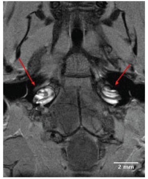
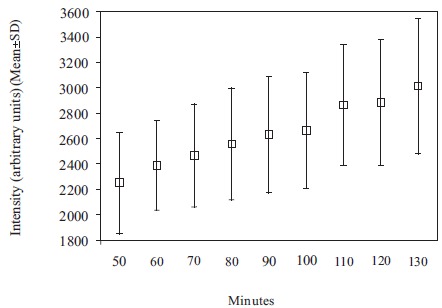
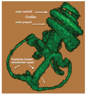
High-resolution 2D and 3D images of the mouse cochlea were acquired within 80 minutes following intravenous injection of Gd. Gd initially permeated the perilymphatic scala tympani and scala vestibuli, and permitted visualization of both cochlear turns from base to apex. The superior, inferior and lateral semicircular canals were subsequently visualized in 3 planes. The membranous endolymphatic labyrinth was impermeable to intravenously injected Gd, and thus showed no apparent uptake of Gd at 9.4T.

CONCLUSION

The 9.4T field strength MRI permitted acquisition of high resolution images of anatomical and physiological features of the normal, wild type mouse perilymphatic inner ear in vivo, and provided further evidence that the endolymphatic system is impermeable to intravenously injected Gd.

摘要

目的

先前在4.7特斯拉场强下对哺乳动物内耳进行的体内实验性磁共振成像(MRI)研究表明,静脉注射钆(Gd)可穿透外淋巴迷路,但不能穿透内淋巴膜迷路。在本研究中,使用9.4T的高场MRI来观察活体小鼠的前庭 - 耳蜗系统,并确定内淋巴系统是否可渗透钆复合物。

方法

使用配备内径为12 cm、最大梯度强度为600 mT/m的梯度系统、千足线圈(Varian设计)和钆造影剂的9.4T Varian磁体,对正常C57 BL - 6小鼠进行图像采集。

结果

静脉注射钆后80分钟内获得了小鼠耳蜗的高分辨率二维和三维图像。钆最初渗透到外淋巴鼓阶和前庭阶,并能从基部到顶部观察到两个耳蜗螺旋。随后在三个平面上观察到上、下和外侧半规管。静脉注射的钆不能穿透膜性内淋巴迷路,因此在9.4T时未显示出明显的钆摄取。

结论

9.4T场强的MRI能够在体内获取正常野生型小鼠外淋巴内耳解剖和生理特征的高分辨率图像,并进一步证明内淋巴系统对静脉注射的钆是不可渗透的。

https://cdn.ncbi.nlm.nih.gov/pmc/blobs/8a08/3722534/1bf69d69666c/TONIJ-7-27_F3.jpg
https://cdn.ncbi.nlm.nih.gov/pmc/blobs/8a08/3722534/b6627c678053/TONIJ-7-27_F1.jpg
https://cdn.ncbi.nlm.nih.gov/pmc/blobs/8a08/3722534/2b78b51180ec/TONIJ-7-27_F2A.jpg
https://cdn.ncbi.nlm.nih.gov/pmc/blobs/8a08/3722534/6ad1dc191c3c/TONIJ-7-27_F2B.jpg
https://cdn.ncbi.nlm.nih.gov/pmc/blobs/8a08/3722534/1bf69d69666c/TONIJ-7-27_F3.jpg

相似文献

1
MRI evidence of endolymphatic impermeability to the gadolinium molecule in the in vivo mouse inner ear at 9.4 tesla.9.4特斯拉下活体小鼠内耳中钆分子内淋巴渗透性的MRI证据。
Open Neuroimag J. 2013 Jun 28;7:27-31. doi: 10.2174/1874440001307010027. Print 2013.
2
Ultra-high-field (9.4 T) MRI Analysis of Contrast Agent Transport Across the Blood-Perilymph Barrier and Intrastrial Fluid-Blood Barrier in the Mouse Inner Ear.超高场(9.4T)磁共振成像分析造影剂在小鼠内耳血-外淋巴屏障和内淋巴液-血屏障中的转运情况。
Otol Neurotol. 2017 Aug;38(7):1052-1059. doi: 10.1097/MAO.0000000000001458.
3
Experimental Fusion of Contrast Enhanced High-Field Magnetic Resonance Imaging and High-Resolution Micro-Computed Tomography in Imaging the Mouse Inner Ear.对比增强高场磁共振成像与高分辨率微型计算机断层扫描在小鼠内耳成像中的实验融合
Open Neuroimag J. 2015 Jul 31;9:7-12. doi: 10.2174/1874440001509010007. eCollection 2015.
4
[Visualization of endolymphatic hydrops in 3D-FLAIR MRI after intratympanic Gd-DTPA administration in Meniere's disease patients].[梅尼埃病患者鼓室内注射钆喷酸葡胺后3D-FLAIR MRI对内淋巴积水的可视化]
Zhonghua Er Bi Yan Hou Tou Jing Wai Ke Za Zhi. 2013 Aug;48(8):628-33.
5
[Optimization of delivering minimum Gd-DTPA at the posterior upper point on tympanic medial wall and hT2W-3D-FLAIR sequence for detecting endolymphatic hydrops].[鼓膜内侧壁后上方点最小剂量钆喷酸葡胺给药及hT2W-3D-FLAIR序列用于检测内淋巴积水的优化]
Zhonghua Er Bi Yan Hou Tou Jing Wai Ke Za Zhi. 2018 Dec 7;53(12):931-938. doi: 10.3760/cma.j.issn.1673-0860.2018.12.011.
6
Diverse patterns of perilymphatic space enhancement in the rat inner ear after intratympanic injection of two different types of gadolinium: a 9.4-tesla magnetic resonance study.经鼓室内注射两种不同类型钆后大鼠内耳外淋巴间隙强化的多样模式:一项9.4特斯拉磁共振研究
Audiol Neurootol. 2015;20(2):112-116. doi: 10.1159/000368666. Epub 2015 Mar 4.
7
3D MRI of the in vivo vestibulo-cochlea labyrinth during Gd-DTPA-BMA uptake.钆喷酸葡胺 - BMA摄取过程中体内前庭蜗迷路的三维磁共振成像。
Neuroreport. 2003 Sep 15;14(13):1707-12. doi: 10.1097/00001756-200309150-00010.
8
In vivo visualization of endolymphatic hydrops in guinea pigs: magnetic resonance imaging evaluation at 4.7 tesla.
Ann Otol Rhinol Laryngol. 2003 Dec;112(12):1059-65. doi: 10.1177/000348940311201212.
9
Clinical significance of endolymphatic imaging after intratympanic gadolinium injection.鼓室内注射钆后内淋巴成像的临床意义
Acta Otolaryngol Suppl. 2009 Feb(560):9-14. doi: 10.1080/00016480902729801.
10
Magnetic resonance imaging of the inner ear in Meniere's disease.梅尼埃病内耳的磁共振成像
Otolaryngol Clin North Am. 2010 Oct;43(5):1059-80. doi: 10.1016/j.otc.2010.06.001.

引用本文的文献

1
Blood Labyrinth Barrier Waste Clearance as Assessed by GBCAs on MRI: A Scoping Review.基于钆对比剂的MRI评估血脑屏障废物清除:一项范围综述
Laryngoscope Investig Otolaryngol. 2025 Sep 9;10(5):e70240. doi: 10.1002/lio2.70240. eCollection 2025 Oct.
2
MRI With Gadolinium as a Measure of Blood-Labyrinth Barrier Integrity in Patients With Inner Ear Symptoms: A Scoping Review.钆增强磁共振成像用于评估内耳症状患者血迷路屏障完整性:一项范围综述
Front Neurol. 2021 May 20;12:662264. doi: 10.3389/fneur.2021.662264. eCollection 2021.
3
Intravenous Delayed Gadolinium-Enhanced MR Imaging of the Endolymphatic Space: A Methodological Comparative Study.

本文引用的文献

1
Imaging of endolymphatic and perilymphatic fluid after intravenous administration of single-dose gadodiamide.静脉单次注射钆喷酸葡胺后内淋巴液和外淋巴液的影像学表现。
Magn Reson Med Sci. 2012;11(2):145-50. doi: 10.2463/mrms.11.145.
2
Mature middle and inner ears express Chd7 and exhibit distinctive pathologies in a mouse model of CHARGE syndrome.成熟的中耳和内耳表达 Chd7,并在 CHARGE 综合征的小鼠模型中表现出独特的病理学特征。
Hear Res. 2011 Dec;282(1-2):184-95. doi: 10.1016/j.heares.2011.08.005. Epub 2011 Aug 23.
3
Direct entry of gadolinium into the vestibule following intratympanic applications in Guinea pigs and the influence of cochlear implantation.
内淋巴囊间隙的静脉注射延迟钆增强磁共振成像:一项方法学比较研究。
Front Neurol. 2021 Apr 22;12:647296. doi: 10.3389/fneur.2021.647296. eCollection 2021.
4
Experimental Fusion of Contrast Enhanced High-Field Magnetic Resonance Imaging and High-Resolution Micro-Computed Tomography in Imaging the Mouse Inner Ear.对比增强高场磁共振成像与高分辨率微型计算机断层扫描在小鼠内耳成像中的实验融合
Open Neuroimag J. 2015 Jul 31;9:7-12. doi: 10.2174/1874440001509010007. eCollection 2015.
豚鼠鼓室内应用后镫骨内直接进入前庭以及耳蜗植入的影响。
J Assoc Res Otolaryngol. 2011 Dec;12(6):741-51. doi: 10.1007/s10162-011-0280-5. Epub 2011 Jul 19.
4
Differential passage of gadolinium through the mouse inner ear barriers evaluated with 4.7T MRI.用 4.7T MRI 评估钆在小鼠内耳屏障中的差异通透性。
Hear Res. 2010 Jan;259(1-2):36-43. doi: 10.1016/j.heares.2009.09.015. Epub 2009 Oct 7.
5
Defects in vestibular sensory epithelia and innervation in mice with loss of Chd7 function: implications for human CHARGE syndrome.Chd7功能缺失小鼠的前庭感觉上皮和神经支配缺陷:对人类CHARGE综合征的启示
J Comp Neurol. 2007 Oct 10;504(5):519-32. doi: 10.1002/cne.21460.
6
Imaging of the guinea pig cochlea following round window gadolinium application.圆窗应用钆后豚鼠耳蜗的成像
Neuroreport. 2004 Aug 26;15(12):1927-30. doi: 10.1097/00001756-200408260-00019.
7
The role of Pax2 in mouse inner ear development.Pax2在小鼠内耳发育中的作用。
Dev Biol. 2004 Aug 1;272(1):161-75. doi: 10.1016/j.ydbio.2004.04.024.
8
In vivo observation of dynamic perilymph formation using 4.7 T MRI with gadolinium as a tracer.使用钆作为示踪剂,通过4.7 T磁共振成像对动态外淋巴形成进行体内观察。
Acta Otolaryngol. 2003 Oct;123(8):910-5. doi: 10.1080/00016480310000548.
9
3D MRI of the in vivo vestibulo-cochlea labyrinth during Gd-DTPA-BMA uptake.钆喷酸葡胺 - BMA摄取过程中体内前庭蜗迷路的三维磁共振成像。
Neuroreport. 2003 Sep 15;14(13):1707-12. doi: 10.1097/00001756-200309150-00010.
10
Magnetic resonance imaging of the membranous labyrinth during in vivo gadolinium (Gd-DTPA-BMA) uptake in the normal and lesioned cochlea.
Neuroreport. 2000 Dec 18;11(18):3979-83. doi: 10.1097/00001756-200012180-00015.