• 文献检索
  • 文档翻译
  • 深度研究
  • 学术资讯
  • Suppr Zotero 插件Zotero 插件
  • 邀请有礼
  • 套餐&价格
  • 历史记录
应用&插件
Suppr Zotero 插件Zotero 插件浏览器插件Mac 客户端Windows 客户端微信小程序
定价
高级版会员购买积分包购买API积分包
服务
文献检索文档翻译深度研究API 文档MCP 服务
关于我们
关于 Suppr公司介绍联系我们用户协议隐私条款
关注我们

Suppr 超能文献

核心技术专利:CN118964589B侵权必究
粤ICP备2023148730 号-1Suppr @ 2026

文献检索

告别复杂PubMed语法,用中文像聊天一样搜索,搜遍4000万医学文献。AI智能推荐,让科研检索更轻松。

立即免费搜索

文件翻译

保留排版,准确专业,支持PDF/Word/PPT等文件格式,支持 12+语言互译。

免费翻译文档

深度研究

AI帮你快速写综述,25分钟生成高质量综述,智能提取关键信息,辅助科研写作。

立即免费体验

与指导囊泡停泊的活性区物质中的大分子对准突触囊泡大分子。

Alignment of synaptic vesicle macromolecules with the macromolecules in active zone material that direct vesicle docking.

机构信息

Department of Neurobiology, Stanford University School of Medicine, Stanford, California, USA.

出版信息

PLoS One. 2013 Jul 22;8(7):e69410. doi: 10.1371/journal.pone.0069410. Print 2013.

DOI:10.1371/journal.pone.0069410
PMID:23894473
原文链接:https://pmc.ncbi.nlm.nih.gov/articles/PMC3718691/
Abstract

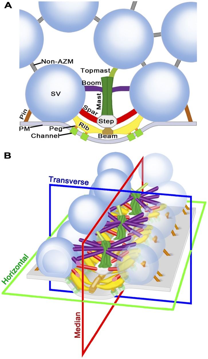
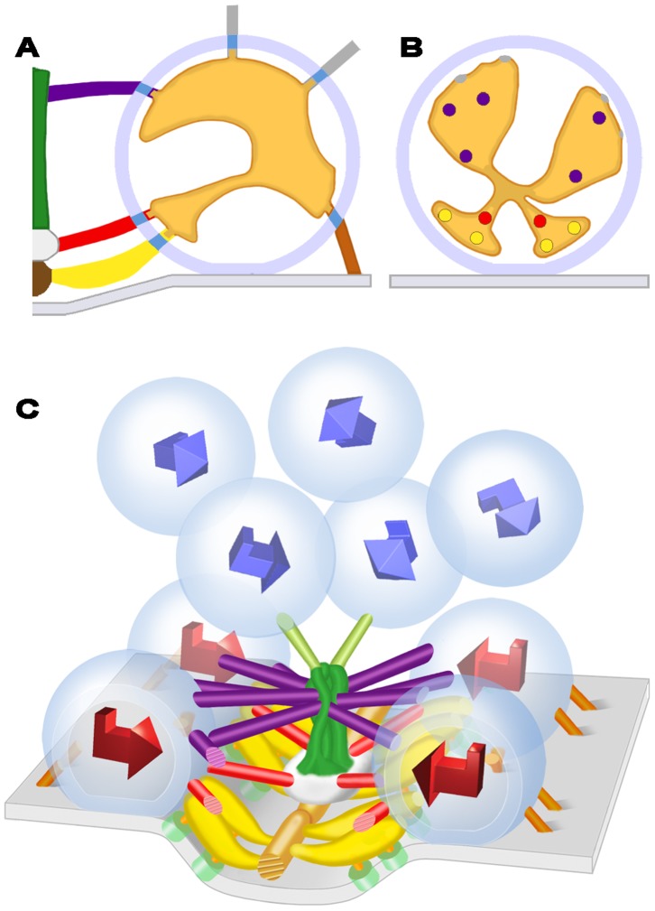
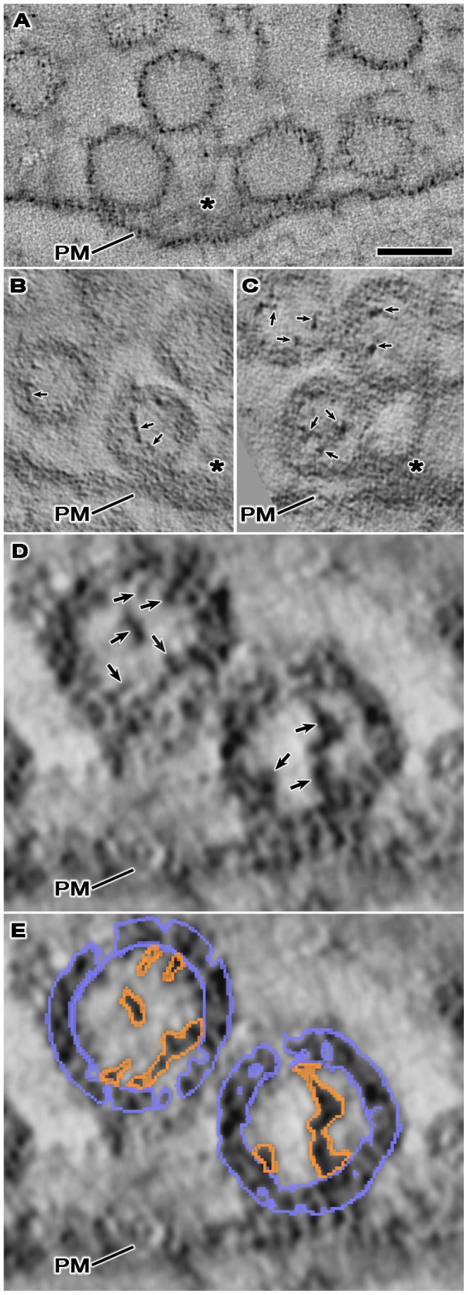
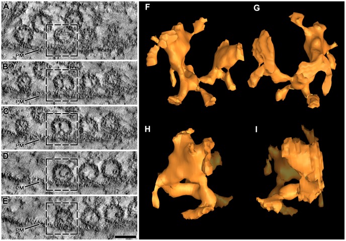
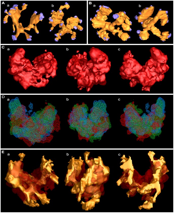
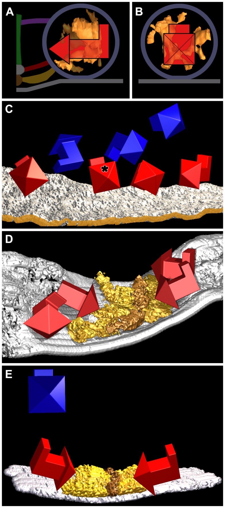
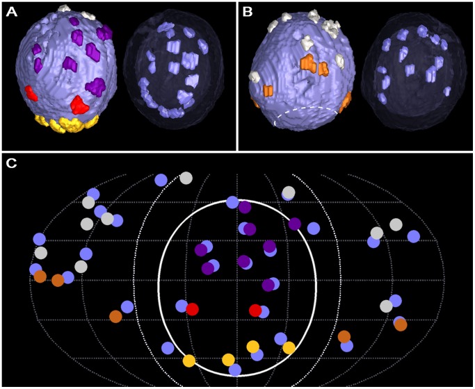
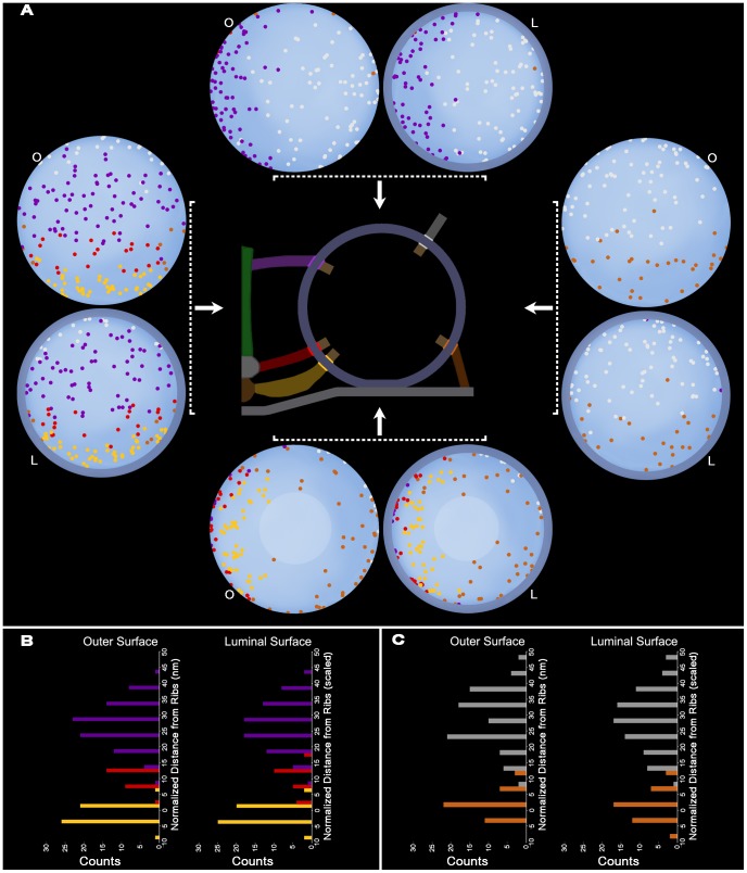
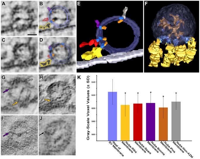
Synaptic vesicles dock at active zones on the presynaptic plasma membrane of a neuron's axon terminals as a precondition for fusing with the membrane and releasing their neurotransmitter to mediate synaptic impulse transmission. Typically, docked vesicles are next to aggregates of plasma membrane-bound macromolecules called active zone material (AZM). Electron tomography on tissue sections from fixed and stained axon terminals of active and resting frog neuromuscular junctions has led to the conclusion that undocked vesicles are directed to and held at the docking sites by the successive formation of stable connections between vesicle membrane proteins and proteins in different classes of AZM macromolecules. Using the same nanometer scale 3D imaging technology on appropriately stained frog neuromuscular junctions, we found that ∼10% of a vesicle's luminal volume is occupied by a radial assembly of elongate macromolecules attached by narrow projections, nubs, to the vesicle membrane at ∼25 sites. The assembly's chiral, bilateral shape is nearly the same vesicle to vesicle, and nubs, at their sites of connection to the vesicle membrane, are linked to macromolecules that span the membrane. For docked vesicles, the orientation of the assembly's shape relative to the AZM and the presynaptic membrane is the same vesicle to vesicle, whereas for undocked vesicles it is not. The connection sites of most nubs on the membrane of docked vesicles are paired with the connection sites of the different classes of AZM macromolecules that regulate docking, and the membrane spanning macromolecules linked to these nubs are also attached to the AZM macromolecules. We conclude that the luminal assembly of macromolecules anchors in a particular arrangement vesicle membrane macromolecules, which contain the proteins that connect the vesicles to AZM macromolecules during docking. Undocked vesicles must move in a way that aligns this arrangement with the AZM macromolecules for docking to proceed.

摘要

突触小泡在神经元轴突末梢的突触前质膜的活性区停靠,作为与膜融合并释放其神经递质以介导突触冲动传递的前提条件。通常,停靠的小泡位于称为活性区物质(AZM)的质膜结合大分子聚集物旁边。对固定和染色的活性和静止蛙运动神经元突触的轴突末梢的组织切片进行电子断层扫描,得出的结论是,未停靠的小泡通过囊泡膜蛋白与不同类别的 AZM 大分子中的蛋白之间稳定连接的连续形成被引导到停靠位点并保持在该位点。在适当染色的蛙运动神经元突触上使用相同的纳米级 3D 成像技术,我们发现小泡内腔体积的约 10%被一组拉长的大分子占据,这些大分子通过狭窄的突起、小结与囊泡膜相连,在约 25 个位点处。该组装体的手性、双侧形状在囊泡之间几乎相同,而在与囊泡膜连接的位点处的小结与跨膜的大分子相连。对于停靠的小泡,组装体形状相对于 AZM 和突触前质膜的取向在囊泡之间是相同的,而对于未停靠的小泡则不是。大多数小结在停靠小泡膜上的连接位点与调节停靠的不同类别的 AZM 大分子的连接位点配对,与这些小结相连的跨膜大分子也与 AZM 大分子相连。我们得出结论,内腔大分子的组装以特定的方式锚定囊泡膜大分子,这些大分子包含将囊泡与 AZM 大分子连接的蛋白质,在停靠过程中。未停靠的小泡必须以一种方式移动,使这种排列与 AZM 大分子对齐,以便进行停靠。

https://cdn.ncbi.nlm.nih.gov/pmc/blobs/25b5/3718691/fb05ae75ed42/pone.0069410.g009.jpg
https://cdn.ncbi.nlm.nih.gov/pmc/blobs/25b5/3718691/029b7179a6ed/pone.0069410.g001.jpg
https://cdn.ncbi.nlm.nih.gov/pmc/blobs/25b5/3718691/9415e2fddf3e/pone.0069410.g002.jpg
https://cdn.ncbi.nlm.nih.gov/pmc/blobs/25b5/3718691/1138d85e80dc/pone.0069410.g003.jpg
https://cdn.ncbi.nlm.nih.gov/pmc/blobs/25b5/3718691/fb7e20c8b905/pone.0069410.g004.jpg
https://cdn.ncbi.nlm.nih.gov/pmc/blobs/25b5/3718691/271f888a6fa4/pone.0069410.g005.jpg
https://cdn.ncbi.nlm.nih.gov/pmc/blobs/25b5/3718691/a7435b30077a/pone.0069410.g006.jpg
https://cdn.ncbi.nlm.nih.gov/pmc/blobs/25b5/3718691/b195f96af692/pone.0069410.g007.jpg
https://cdn.ncbi.nlm.nih.gov/pmc/blobs/25b5/3718691/73065999c330/pone.0069410.g008.jpg
https://cdn.ncbi.nlm.nih.gov/pmc/blobs/25b5/3718691/fb05ae75ed42/pone.0069410.g009.jpg

相似文献

1
Alignment of synaptic vesicle macromolecules with the macromolecules in active zone material that direct vesicle docking.与指导囊泡停泊的活性区物质中的大分子对准突触囊泡大分子。
PLoS One. 2013 Jul 22;8(7):e69410. doi: 10.1371/journal.pone.0069410. Print 2013.
2
Regulation of synaptic vesicle docking by different classes of macromolecules in active zone material.不同种类的大分子在活性区物质中对突触小泡 docking 的调节。
PLoS One. 2012;7(3):e33333. doi: 10.1371/journal.pone.0033333. Epub 2012 Mar 16.
3
Macromolecular connections of active zone material to docked synaptic vesicles and presynaptic membrane at neuromuscular junctions of mouse.在小鼠神经肌肉接头处,活性区物质与对接的突触小泡和突触前膜的大分子连接。
J Comp Neurol. 2009 Apr 10;513(5):457-68. doi: 10.1002/cne.21975.
4
Active Zone Material-Directed Orientation, Docking, and Fusion of Dense Core Vesicles Alongside Synaptic Vesicles at Neuromuscular Junctions.活性区物质导向的致密核心囊泡在神经肌肉接头处与突触囊泡一起的定向、对接和融合。
Front Neuroanat. 2018 Sep 13;12:72. doi: 10.3389/fnana.2018.00072. eCollection 2018.
5
Synaptic Vesicles Having Large Contact Areas with the Presynaptic Membrane are Preferentially Hemifused at Active Zones of Frog Neuromuscular Junctions Fixed during Synaptic Activity.在突触活动期间固定的蛙类神经肌肉接头的活性区,与突触前膜具有大接触面积的突触小泡优先发生半融合。
Int J Mol Sci. 2019 May 31;20(11):2692. doi: 10.3390/ijms20112692.
6
The structure and function of 'active zone material' at synapses.突触处“活性区物质”的结构与功能。
Philos Trans R Soc Lond B Biol Sci. 2015 Jul 5;370(1672). doi: 10.1098/rstb.2014.0189.
7
A stochastic model of active zone material mediated synaptic vesicle docking and priming at resting active zones.一个活跃区物质介导的突触囊泡停泊和引发的随机模型,位于静止的活跃区。
Sci Rep. 2017 Mar 21;7(1):278. doi: 10.1038/s41598-017-00360-z.
8
Hypothesis Relating the Structure, Biochemistry and Function of Active Zone Material Macromolecules at a Neuromuscular Junction.关于神经肌肉接头处活性区物质大分子的结构、生物化学与功能的假说
Front Synaptic Neurosci. 2022 Jan 5;13:798225. doi: 10.3389/fnsyn.2021.798225. eCollection 2021.
9
Synapsin II and calcium regulate vesicle docking and the cross-talk between vesicle pools at the mouse motor terminals.突触结合蛋白II和钙调节小鼠运动终板处的囊泡对接以及囊泡池之间的相互作用。
J Physiol. 2008 Oct 1;586(19):4649-73. doi: 10.1113/jphysiol.2008.154666. Epub 2008 Jul 31.
10
Variable priming of a docked synaptic vesicle.对接突触小泡的可变引发
Proc Natl Acad Sci U S A. 2016 Feb 23;113(8):E1098-107. doi: 10.1073/pnas.1523054113. Epub 2016 Feb 8.

引用本文的文献

1
Synaptic vesicle glycoprotein 2C enhances vesicular storage of dopamine and counters dopaminergic toxicity.突触囊泡糖蛋白 2C 增强多巴胺囊泡储存,并对抗多巴胺能毒性。
Eur J Neurosci. 2024 May;59(10):2483-2501. doi: 10.1111/ejn.16311. Epub 2024 Mar 26.
2
Microphysiological Modeling of the Structure and Function of Neuromuscular Transmitter Release Sites.神经肌肉递质释放位点结构与功能的微生理建模
Front Synaptic Neurosci. 2022 Jun 13;14:917285. doi: 10.3389/fnsyn.2022.917285. eCollection 2022.
3
Brivaracetam and Levetiracetam Suppress Astroglial L-Glutamate Release through Hemichannel via Inhibition of Synaptic Vesicle Protein.

本文引用的文献

1
Regulation of synaptic vesicle docking by different classes of macromolecules in active zone material.不同种类的大分子在活性区物质中对突触小泡 docking 的调节。
PLoS One. 2012;7(3):e33333. doi: 10.1371/journal.pone.0033333. Epub 2012 Mar 16.
2
Protein quantification at the single vesicle level reveals that a subset of synaptic vesicle proteins are trafficked with high precision.在单个囊泡水平进行蛋白质定量分析表明,一部分突触囊泡蛋白以高精度运输。
J Neurosci. 2011 Jan 26;31(4):1461-70. doi: 10.1523/JNEUROSCI.3805-10.2011.
3
Structure parameters of synaptic vesicles quantified by small-angle x-ray scattering.
布瓦西坦和左乙拉西坦通过抑制突触囊泡蛋白经由缝隙连接抑制星形胶质细胞 L-谷氨酸释放。
Int J Mol Sci. 2022 Apr 19;23(9):4473. doi: 10.3390/ijms23094473.
4
Hypothesis Relating the Structure, Biochemistry and Function of Active Zone Material Macromolecules at a Neuromuscular Junction.关于神经肌肉接头处活性区物质大分子的结构、生物化学与功能的假说
Front Synaptic Neurosci. 2022 Jan 5;13:798225. doi: 10.3389/fnsyn.2021.798225. eCollection 2021.
5
Promising drug targets and associated therapeutic interventions in Parkinson's disease.帕金森病中有前景的药物靶点及相关治疗干预措施。
Neural Regen Res. 2021 Sep;16(9):1730-1739. doi: 10.4103/1673-5374.306066.
6
Impairment Mechanisms and Intervention Approaches for Aged Human Neuromuscular Junctions.老年人神经肌肉接头的损伤机制与干预方法
Front Mol Neurosci. 2020 Nov 16;13:568426. doi: 10.3389/fnmol.2020.568426. eCollection 2020.
7
The Decade of Super-Resolution Microscopy of the Presynapse.突触超分辨率显微镜十年
Front Synaptic Neurosci. 2020 Aug 11;12:32. doi: 10.3389/fnsyn.2020.00032. eCollection 2020.
8
The Synaptic Vesicle Glycoprotein 2: Structure, Function, and Disease Relevance.突触囊泡糖蛋白 2:结构、功能和疾病相关性。
ACS Chem Neurosci. 2019 Sep 18;10(9):3927-3938. doi: 10.1021/acschemneuro.9b00351. Epub 2019 Aug 23.
9
Synaptic Vesicles Having Large Contact Areas with the Presynaptic Membrane are Preferentially Hemifused at Active Zones of Frog Neuromuscular Junctions Fixed during Synaptic Activity.在突触活动期间固定的蛙类神经肌肉接头的活性区,与突触前膜具有大接触面积的突触小泡优先发生半融合。
Int J Mol Sci. 2019 May 31;20(11):2692. doi: 10.3390/ijms20112692.
10
Active Zone Material-Directed Orientation, Docking, and Fusion of Dense Core Vesicles Alongside Synaptic Vesicles at Neuromuscular Junctions.活性区物质导向的致密核心囊泡在神经肌肉接头处与突触囊泡一起的定向、对接和融合。
Front Neuroanat. 2018 Sep 13;12:72. doi: 10.3389/fnana.2018.00072. eCollection 2018.
小角度 X 射线散射定量分析突触小泡的结构参数。
Biophys J. 2010 Apr 7;98(7):1200-8. doi: 10.1016/j.bpj.2009.12.4278.
4
Quantitative analysis of the native presynaptic cytomatrix by cryoelectron tomography.通过冷冻电镜断层扫描对天然突触前细胞外基质进行定量分析。
J Cell Biol. 2010 Jan 11;188(1):145-56. doi: 10.1083/jcb.200908082.
5
A common molecular basis for membrane docking and functional priming of synaptic vesicles.突触小泡膜对接和功能启动的共同分子基础。
Eur J Neurosci. 2009 Jul;30(1):49-56. doi: 10.1111/j.1460-9568.2009.06811.x. Epub 2009 Jun 25.
6
Macromolecular connections of active zone material to docked synaptic vesicles and presynaptic membrane at neuromuscular junctions of mouse.在小鼠神经肌肉接头处,活性区物质与对接的突触小泡和突触前膜的大分子连接。
J Comp Neurol. 2009 Apr 10;513(5):457-68. doi: 10.1002/cne.21975.
7
Mechanics of membrane fusion.膜融合机制。
Nat Struct Mol Biol. 2008 Jul;15(7):675-83. doi: 10.1038/nsmb.1455.
8
Conical electron tomography of a chemical synapse: polyhedral cages dock vesicles to the active zone.化学突触的锥形电子断层扫描:多面体笼将囊泡对接至活性区。
J Neurosci. 2008 Apr 16;28(16):4151-60. doi: 10.1523/JNEUROSCI.4639-07.2008.
9
Organization of the core structure of the postsynaptic density.突触后致密区核心结构的组织
Proc Natl Acad Sci U S A. 2008 Mar 18;105(11):4453-8. doi: 10.1073/pnas.0800897105. Epub 2008 Mar 7.
10
The combination of chemical fixation procedures with high pressure freezing and freeze substitution preserves highly labile tissue ultrastructure for electron tomography applications.化学固定程序与高压冷冻和冷冻置换相结合,可为电子断层扫描应用保留高度不稳定的组织超微结构。
J Struct Biol. 2008 Mar;161(3):359-71. doi: 10.1016/j.jsb.2007.09.002. Epub 2007 Sep 14.