Tabuchi Sadaharu, Nakajima Sadao
Department of Neurosurgery, Tottori Prefectural Central Hospital, Tottori, Japan.
Case Rep Oncol. 2013 Jul 6;6(2):362-6. doi: 10.1159/000353929. Print 2013 May.
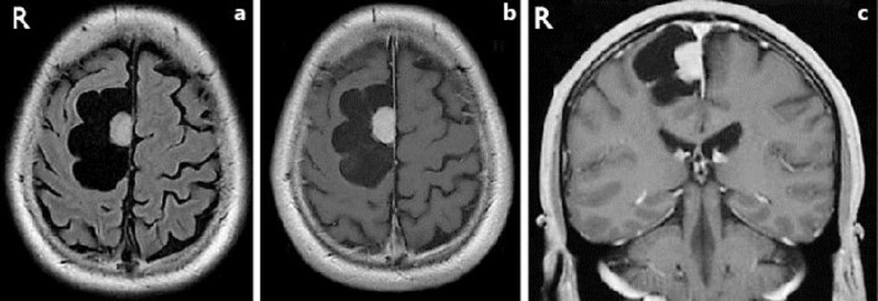
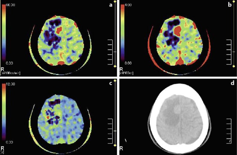
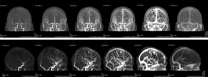
We present a case of cystic falx meningioma. Cystic meningioma is rare and not easy to diagnose preoperatively; it is often misdiagnosed as other tumors, including glial or metastatic tumors with cystic or necrotic changes. This study showed the potential impact of 320-row computed tomography (CT) on image-based diagnostic evaluation of cystic meningioma with special attention to the novel techniques of 4-dimensional CT angiography (4D-CTA) and CT whole-brain perfusion (CTP). 4D-CTA showed the arterial supply feeding the tumor and late enhancement of the tumor nodule, similar to that seen in meningioma by conventional angiography. CTP showed that the tumor had a higher cerebral blood flow and cerebral blood volume and a longer mean transit time than adjacent brain tissue. These findings were consistent with meningioma and reinforced the other imaging findings, resulting in the correct preoperative diagnosis. The new techniques available for 320-row CT can potentially be used to improve differential diagnosis and preoperative assessment of cystic tumors with nodules.
我们报告一例囊性大脑镰脑膜瘤病例。囊性脑膜瘤较为罕见,术前不易诊断;它常被误诊为其他肿瘤,包括伴有囊性或坏死改变的神经胶质瘤或转移瘤。本研究显示了320排计算机断层扫描(CT)对基于图像的囊性脑膜瘤诊断评估的潜在影响,特别关注四维CT血管造影(4D-CTA)和CT全脑灌注(CTP)等新技术。4D-CTA显示了为肿瘤供血的动脉供应以及肿瘤结节的延迟强化,这与传统血管造影所见的脑膜瘤相似。CTP显示肿瘤的脑血流量和脑血容量高于相邻脑组织,平均通过时间更长。这些发现与脑膜瘤一致,并强化了其他影像学表现,从而实现了正确的术前诊断。320排CT可用的新技术有可能用于改善对伴有结节的囊性肿瘤的鉴别诊断和术前评估。