Keller Kate E, Yang Yong-Feng, Sun Ying Ying, Sykes Renee, Acott Ted S, Wirtz Mary K
Casey Eye Institute, Oregon Health & Science University, 3181 SW Sam Jackson Park Rd, Portland, OR 97239, USA.
Mol Vis. 2013 Jul 25;19:1639-55. Print 2013.
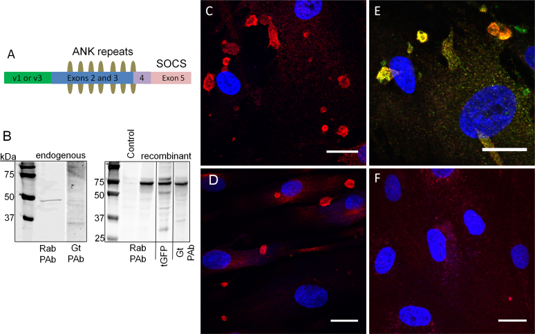
Ankyrin repeat and suppressor of cytokine signaling (SOCS) box containing protein-10 (ASB10) was recently identified as a gene that causes primary open-angle glaucoma. Here, we investigated endogenous ASB10 protein expression in human trabecular meshwork (HTM) cells to provide the first clues to the biologic function of this protein.
Primary HTM cells were cultured and immunostained with anti-ASB10 and various biomarkers of the ubiquitin-mediated proteasomal and autophagy-lysosomal degradation pathways. Cells were imaged with confocal and high-resolution structured illumination microscopy. Colocalization was quantified using Imaris Bitplane software, which generated a Pearson's correlation coefficient value. Coimmunoprecipitation of ASB10-transfected cells was performed.
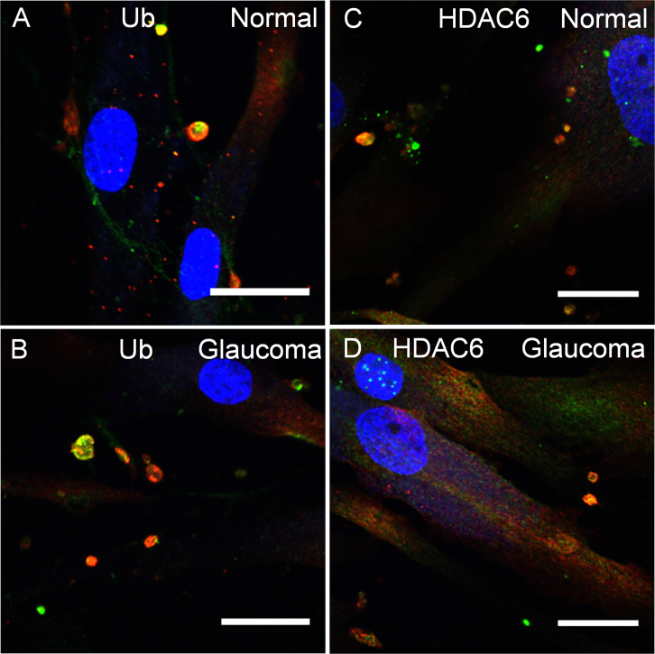
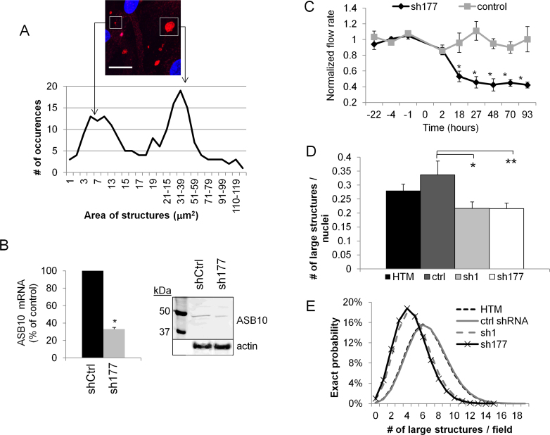
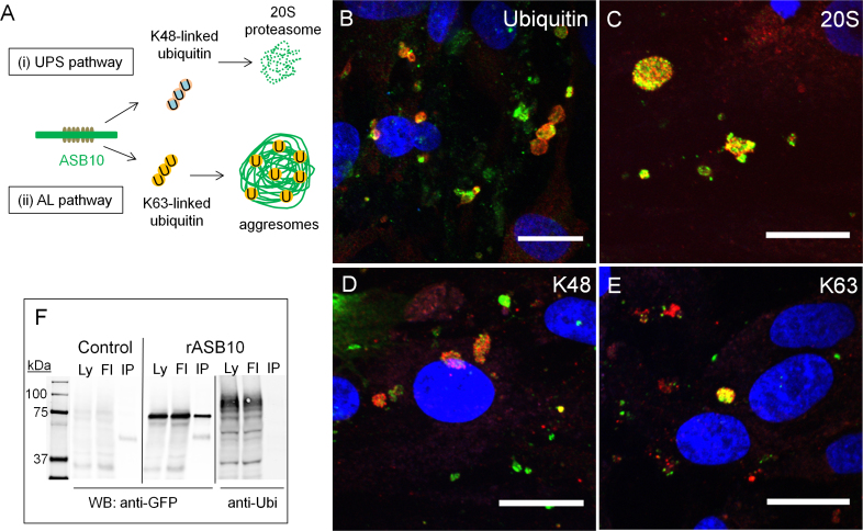
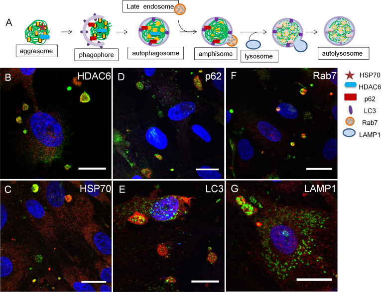
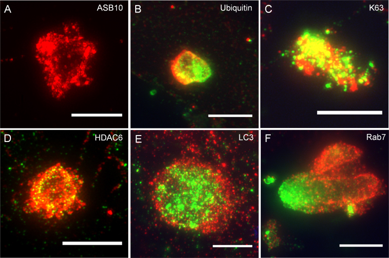
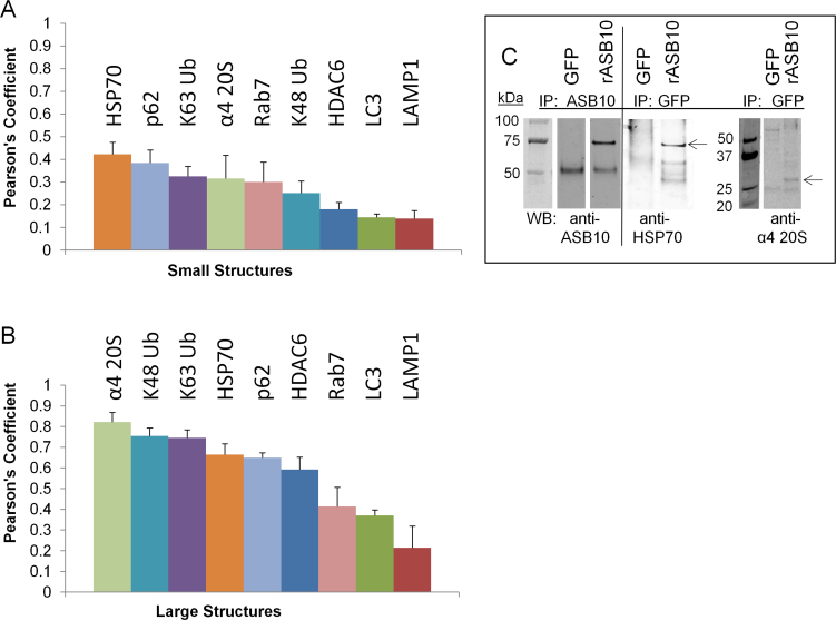
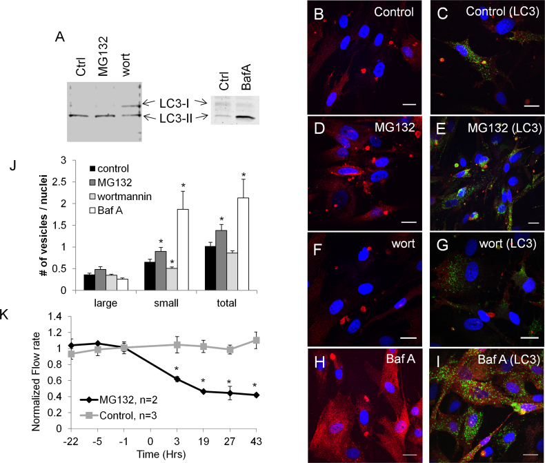
Immunofluorescence and confocal analysis showed that ASB10 was localized in intracellular structures in HTM cells. Two populations were observed: small, spherical vesicles and larger, less abundant structures. In the ASB10-silenced cells, the number of large structures was significantly decreased. ASB10 partially colocalized with biomarkers of the ubiquitin-mediated proteasomal pathway including ubiquitin and the α4 subunit of the 20S proteasome. However, ASB10 itself was not ubiquitinated. ASB10 also colocalized with numerous biomarkers of specific autophagic structures: aggresomes (histone deacetylase 6 [HDAC6] and heat shock protein 70 [HSP70]), autophagosomes (light chain 3 [LC3] and p62), amphisomes (Rab7), and lysosomes (lysosomal-associated membrane protein 1 [LAMP1]). Pearson coefficients indicated strong colocalization of large ASB10-stained structures with the α4 subunit of the 20S proteasome, K48 and K63-linked ubiquitin antibodies, p62, HSP70, and HDAC6 (Pearson's range, 0.59-0.82). Coimmunoprecipitation assays showed a positive interaction of ASB10 with HSP70 and with the α4 subunit of the 20S proteasome. Super-resolution structured illumination confocal microscopy suggested that the smaller ASB10-stained vesicles aggregated into the larger structures, which resembled aggresome-like induced structures. Treatment of HTM cells with an autophagy activator (MG132) or inhibitors (wortmannin, bafilomycin A1) significantly increased and decreased the number of small ASB10-stained vesicles, respectively. No discernible differences in the colocalization of large ASB10-stained structures with ubiquitin or HDAC6 were observed between dermal fibroblasts derived from a normal individual and a patient with primary open-angle glaucoma carrying a synonymous ASB10 mutation.
Our evidence suggests that ASB10 may play a role in ubiquitin-mediated degradation pathways in TM cells.
锚蛋白重复序列和细胞因子信号转导抑制因子(SOCS)盒包含蛋白10(ASB10)最近被鉴定为导致原发性开角型青光眼的基因。在此,我们研究了人小梁网(HTM)细胞中内源性ASB10蛋白的表达,以提供有关该蛋白生物学功能的首个线索。
培养原代HTM细胞,并用抗ASB10抗体以及泛素介导的蛋白酶体和自噬-溶酶体降解途径的各种生物标志物进行免疫染色。使用共聚焦和高分辨率结构光照显微镜对细胞进行成像。使用Imaris Bitplane软件对共定位进行定量分析,该软件生成皮尔逊相关系数值。对转染了ASB10的细胞进行免疫共沉淀。
免疫荧光和共聚焦分析表明,ASB10定位于HTM细胞的细胞内结构中。观察到两种类型:小的球形囊泡和较大的、数量较少的结构。在ASB10沉默的细胞中,大结构的数量显著减少。ASB10与泛素介导的蛋白酶体途径的生物标志物部分共定位,包括泛素和20S蛋白酶体的α4亚基。然而,ASB10本身未被泛素化。ASB10还与特定自噬结构的多种生物标志物共定位:聚集体(组蛋白去乙酰化酶6 [HDAC6]和热休克蛋白70 [HSP70])、自噬体(微管相关蛋白轻链3 [LC3]和p62)、双膜泡体(Rab7)和溶酶体(溶酶体相关膜蛋白1 [LAMP1])。皮尔逊系数表明,大的ASB10染色结构与20S蛋白酶体的α4亚基、K48和K63连接的泛素抗体、p62、HSP70和HDAC6有很强的共定位(皮尔逊系数范围为0.59 - 0.82)。免疫共沉淀试验表明,ASB10与HSP70以及20S蛋白酶体的α4亚基有阳性相互作用。超分辨率结构光照共聚焦显微镜显示,较小的ASB10染色囊泡聚集形成较大的结构,类似于聚集体样诱导结构。用自噬激活剂(MG132)或抑制剂(渥曼青霉素、巴弗洛霉素A1)处理HTM细胞,分别显著增加和减少了小的ASB10染色囊泡的数量。在来自正常个体的真皮成纤维细胞和携带同义ASB10突变的原发性开角型青光眼患者的真皮成纤维细胞之间,未观察到ASB10染色大结构与泛素或HDAC6共定位的明显差异。
我们的证据表明,ASB10可能在小梁网细胞的泛素介导的降解途径中起作用。