• 文献检索
  • 文档翻译
  • 深度研究
  • 学术资讯
  • Suppr Zotero 插件Zotero 插件
  • 邀请有礼
  • 套餐&价格
  • 历史记录
应用&插件
Suppr Zotero 插件Zotero 插件浏览器插件Mac 客户端Windows 客户端微信小程序
定价
高级版会员购买积分包购买API积分包
服务
文献检索文档翻译深度研究API 文档MCP 服务
关于我们
关于 Suppr公司介绍联系我们用户协议隐私条款
关注我们

Suppr 超能文献

核心技术专利:CN118964589B侵权必究
粤ICP备2023148730 号-1Suppr @ 2026

文献检索

告别复杂PubMed语法,用中文像聊天一样搜索,搜遍4000万医学文献。AI智能推荐,让科研检索更轻松。

立即免费搜索

文件翻译

保留排版,准确专业,支持PDF/Word/PPT等文件格式,支持 12+语言互译。

免费翻译文档

深度研究

AI帮你快速写综述,25分钟生成高质量综述,智能提取关键信息,辅助科研写作。

立即免费体验

经食管超声心动图用于部分无顶冠状静脉窦的冠状静脉窦成像。

Transesophageal echocardiography for coronary sinus imaging in partially unroofed coronary sinus.

作者信息

Sun Ting, Fei Hong-Wen, Huang Huan-Lei, Chen Ou-Di, Zheng Zhi-Chao, Zhang Cao-Jin, Hou Yue-Shuang

机构信息

Department of Cardiology, Guangdong Provincial Cardiovascular Institute, Guangdong General Hospital, Guangdong Academy of Medical Science, Guangzhou, China.

出版信息

Echocardiography. 2014;31(1):74-82. doi: 10.1111/echo.12302. Epub 2013 Jul 30.

DOI:10.1111/echo.12302
PMID:23909710
原文链接:https://pmc.ncbi.nlm.nih.gov/articles/PMC4232913/
Abstract

BACKGROUND

Partially unroofed coronary sinus (PUCS) is a rare congenital cardiac anomaly and prone to be misdiagnosed. The purpose of this study was to explore the value of transesophageal echocardiography (TEE) in CS imaging for the detection of PUCS and to develop a special two-dimensional TEE-based en face view of CS.

METHODS

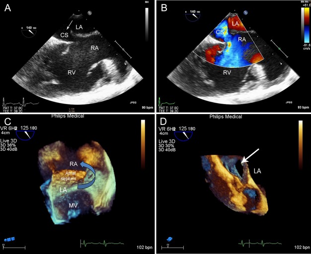
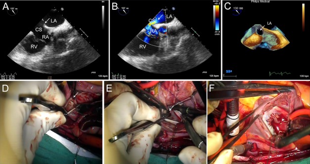
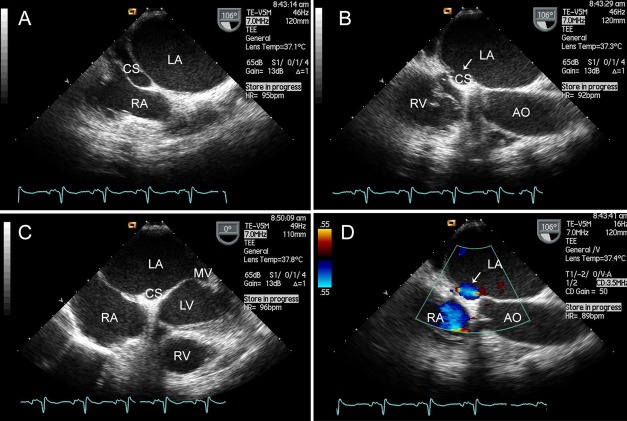
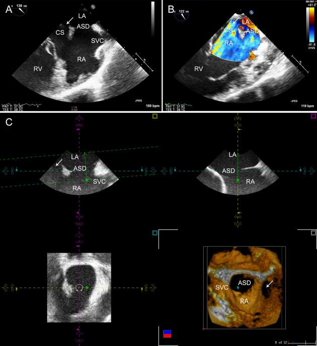
Twenty adult patients with suspected PUCS, showing a dilated coronary sinus and an enlarged right heart on transthoracic echocardiography (TTE), underwent TEE examination. In the mid-esophageal plane and close to an angle of 120°, the en face view of the CS successfully imaged the roof of the CS, which was beyond the realm of the atrial septum, and the interatrial septum was obtained simultaneously in the same view. Meanwhile, the 3D zoom mode could clearly display the comprehensive volume image and the adjacent structures of the PUCS. The results of TEE were compared with the findings of surgery or catheterization.

RESULTS

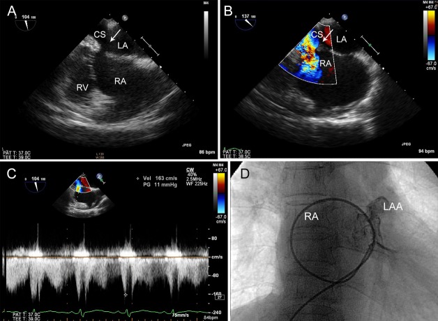
En face view of the CS was obtained successfully by 2DTEE in 20 patients. In addition, 3DTEE was used for imaging of PUCS in 11 of the 20 patients. PUCS was ultimately confirmed in 13 patients either by surgery or catheterization. The TEE for PUCS diagnosis was consistent with the surgical findings.

CONCLUSION

Transesophageal echocardiography can be successfully applied to obtain the comprehensive view of CS and its surrounding structures. The en face view of CS provided by 2DTEE may be helpful in better understanding PUCS and discriminating it from associated atrial septal defects.

摘要

背景

部分无顶冠状静脉窦(PUCS)是一种罕见的先天性心脏异常,容易被误诊。本研究的目的是探讨经食管超声心动图(TEE)在冠状静脉窦(CS)成像中检测PUCS的价值,并开发一种基于二维TEE的CS特殊正面视图。

方法

20例经胸超声心动图(TTE)显示冠状静脉窦扩张和右心增大、疑似PUCS的成年患者接受了TEE检查。在食管中段平面且接近120°角时,CS的正面视图成功成像了CS的顶部,该顶部超出了房间隔的范围,并且在同一视图中同时获得了房间隔。同时,三维缩放模式可以清晰显示PUCS的整体容积图像及其相邻结构。将TEE的结果与手术或导管检查的结果进行比较。

结果

20例患者通过二维TEE成功获得了CS的正面视图。此外,20例患者中的11例使用三维TEE对PUCS进行成像。最终13例患者经手术或导管检查确诊为PUCS。TEE对PUCS的诊断与手术结果一致。

结论

经食管超声心动图可以成功应用于获取CS及其周围结构的全貌。二维TEE提供的CS正面视图可能有助于更好地理解PUCS并将其与相关的房间隔缺损区分开来。

https://cdn.ncbi.nlm.nih.gov/pmc/blobs/ce3c/4232913/a33e958cd11a/echo0031-0074-f5.jpg
https://cdn.ncbi.nlm.nih.gov/pmc/blobs/ce3c/4232913/1f10f8e643b2/echo0031-0074-f1.jpg
https://cdn.ncbi.nlm.nih.gov/pmc/blobs/ce3c/4232913/c25b0f2a3e8f/echo0031-0074-f2.jpg
https://cdn.ncbi.nlm.nih.gov/pmc/blobs/ce3c/4232913/bc5f43500e33/echo0031-0074-f3.jpg
https://cdn.ncbi.nlm.nih.gov/pmc/blobs/ce3c/4232913/43ffb2b0e812/echo0031-0074-f4.jpg
https://cdn.ncbi.nlm.nih.gov/pmc/blobs/ce3c/4232913/a33e958cd11a/echo0031-0074-f5.jpg

相似文献

1
Transesophageal echocardiography for coronary sinus imaging in partially unroofed coronary sinus.经食管超声心动图用于部分无顶冠状静脉窦的冠状静脉窦成像。
Echocardiography. 2014;31(1):74-82. doi: 10.1111/echo.12302. Epub 2013 Jul 30.
2
Utility of three-dimensional transesophageal echocardiography for diagnosis of unroofed coronary sinus.三维经食管超声心动图在诊断无顶冠状静脉窦中的应用
J Med Ultrason (2001). 2016 Jan;43(1):91-4. doi: 10.1007/s10396-015-0648-y. Epub 2015 Jul 7.
3
Partially unroofed coronary sinus diagnosed by real-time three-dimensional transesophageal echocardiography after operation of secundum atrial septal defect.继发孔型房间隔缺损术后经实时三维经食管超声心动图诊断的部分无顶冠状静脉窦
Int J Cardiovasc Imaging. 2015 Jan;31(1):45-6. doi: 10.1007/s10554-014-0528-3. Epub 2014 Sep 10.
4
Coronary sinus septal defect (unroofed coronary sinus): echocardiographic diagnosis and surgical treatment.冠状窦间隔缺损(未覆盖的冠状窦):超声心动图诊断与手术治疗。
Int J Cardiol. 2013 Sep 30;168(2):1258-63. doi: 10.1016/j.ijcard.2012.11.113. Epub 2012 Dec 23.
5
A rare case of unroofed coronary sinus with a secundum atrial septal defect incidentally detected by computed tomography.计算机断层扫描偶然发现的1例罕见的无顶冠状静脉窦合并继发孔型房间隔缺损。
J Cardiovasc Comput Tomogr. 2020 Nov-Dec;14(6):e105-e106. doi: 10.1016/j.jcct.2019.03.001. Epub 2019 Mar 13.
6
Missed unroofed coronary sinus.未发现冠状静脉窦未闭。
Echocardiography. 2019 Mar;36(3):613-614. doi: 10.1111/echo.14274. Epub 2019 Feb 8.
7
Evaluation of Unroofed Coronary Sinus Syndrome Using Cardiovascular CT Angiography: An Observational Study.应用心血管 CT 血管造影术评估冠状窦未闭综合征:一项观察性研究。
AJR Am J Roentgenol. 2018 Aug;211(2):314-320. doi: 10.2214/AJR.17.19128. Epub 2018 Jun 27.
8
Coronary sinus atrial septal defects in adults over the past 20 years at new Tokyo hospital: case series.20 年来在新东京医院治疗的成年人冠状窦房间隔缺损:病例系列。
J Cardiothorac Surg. 2021 May 29;16(1):150. doi: 10.1186/s13019-021-01522-x.
9
Usefulness of real-time three-dimensional trans-oesophageal echocardiography for detection of isolated unroofed coronary sinus.实时三维经食管超声心动图在检测孤立性冠状静脉窦未闭中的应用价值
Cardiol Young. 2015 Jan;25(1):181-3. doi: 10.1017/S1047951113002400. Epub 2014 Feb 19.
10
Incremental Value of Live/Real Time Three-Dimensional over Two-Dimensional Transesophageal Echocardiography in the Assessment of Atrial Septal Pouch.实时三维经食管超声心动图相对于二维经食管超声心动图在评估房间隔囊袋方面的增量价值。
Echocardiography. 2015 Dec;32(12):1858-67. doi: 10.1111/echo.13111. Epub 2015 Nov 11.

引用本文的文献

1
Unroofed coronary sinus, left-sided superior vena cava and mitral insufficiency: A case report and review of the literature.无顶冠状静脉窦、左侧上腔静脉与二尖瓣关闭不全:一例病例报告并文献复习
World J Cardiol. 2024 Oct 26;16(10):595-603. doi: 10.4330/wjc.v16.i10.595.
2
Agitated saline test as a simple but reliable method of intraoperative diagnosis and evaluation of unroofed coronary sinus.生理盐水激发试验作为一种简单但可靠的术中诊断及评估无顶冠状静脉窦的方法。
JA Clin Rep. 2024 Apr 25;10(1):28. doi: 10.1186/s40981-024-00709-0.
3
The value of cardiac CT in the diagnosis of unroofed coronary sinus syndrome.

本文引用的文献

1
Transcatheter treatment of unroofed coronary sinus.经导管治疗冠状窦顶未覆盖。
Catheter Cardiovasc Interv. 2013 Apr;81(5):849-52. doi: 10.1002/ccd.24519. Epub 2012 Sep 7.
2
Partially unroofed coronary sinus: MDCT and MRI findings.部分心外膜冠状窦:MDCT 和 MRI 表现。
AJR Am J Roentgenol. 2010 Nov;195(5):W331-6. doi: 10.2214/AJR.09.3689.
3
Unroofing the diagnosis.揭顶诊断。
心脏 CT 在诊断冠状窦间隔缺损综合征中的价值。
BMC Cardiovasc Disord. 2022 Dec 2;22(1):516. doi: 10.1186/s12872-022-02966-2.
4
Unroofed Coronary Sinus in a Patient With Prior Surgical Closure of Secundum Atrial Septal Defect.一名曾接受继发孔房间隔缺损外科封堵术患者的无顶冠状静脉窦
Cureus. 2021 Nov 8;13(11):e19350. doi: 10.7759/cureus.19350. eCollection 2021 Nov.
5
A Rare Congenital Cardiac Defect: Isolated Coronary Sinus Septal Defect and the Challenges of Diagnosis in a Child.一种罕见的先天性心脏缺陷:孤立性冠状静脉窦间隔缺损及儿童诊断面临的挑战
CASE (Phila). 2021 Aug 20;5(5):286-291. doi: 10.1016/j.case.2021.07.005. eCollection 2021 Oct.
6
Surgical repair of unroofed coronary sinus with severe mitral regurgitation in an elderly patient.老年患者无顶冠状静脉窦合并严重二尖瓣反流的外科修复术。
J Cardiol Cases. 2018 Oct 22;19(1):29-32. doi: 10.1016/j.jccase.2018.09.002. eCollection 2019 Jan.
7
Post Mitral Valve Replacement: Paravalvular Leak or Unroofed Coronary Sinus?二尖瓣置换术后:瓣周漏还是冠状静脉窦未闭?
CASE (Phila). 2017 Oct 19;1(6):227-229. doi: 10.1016/j.case.2017.07.009. eCollection 2017 Dec.
8
Utility of three-dimensional transesophageal echocardiography for diagnosis of unroofed coronary sinus.三维经食管超声心动图在诊断无顶冠状静脉窦中的应用
J Med Ultrason (2001). 2016 Jan;43(1):91-4. doi: 10.1007/s10396-015-0648-y. Epub 2015 Jul 7.
Int J Cardiovasc Imaging. 2010 Dec;26(8):841-2. doi: 10.1007/s10554-010-9667-3. Epub 2010 Jul 8.
4
Images in cardiovascular medicine. Rare case of an unroofed coronary sinus: diagnosis by multidetector computed tomography.心血管医学影像。罕见的无顶冠状静脉窦病例:多层螺旋计算机断层扫描诊断
Circulation. 2009 Apr 28;119(16):e518-20. doi: 10.1161/CIRCULATIONAHA.107.707018.
5
Unroofed coronary sinus with persistent left superior vena cava assessed by 3D echocardiography.三维超声心动图评估无顶冠状静脉窦合并永存左上腔静脉
Echocardiography. 2008 Jul;25(6):666-7. doi: 10.1111/j.1540-8175.2008.00649.x. Epub 2008 Mar 27.
6
Images in cardiovascular medicine. Partially unroofed coronary sinus.心血管医学影像。部分无顶冠状静脉窦。
Circulation. 2007 Oct 9;116(15):e373. doi: 10.1161/CIRCULATIONAHA.107.717942.
7
Assessment of surgically unroofed coronary sinus by live/real time three-dimensional transthoracic echocardiography.经胸实时三维超声心动图对手术开窗冠状静脉窦的评估
Echocardiography. 2007 Jan;24(1):74-6. doi: 10.1111/j.1540-8175.2006.00359.x.
8
Percutaneous closure of a persistent left superior vena cava connected to the left atrium.经皮闭合与左心房相连的永存左上腔静脉。
Int J Cardiol. 2006 Jan 26;106(3):365-6. doi: 10.1016/j.ijcard.2005.02.015.
9
Electrical disconnection of the coronary sinus by radiofrequency catheter ablation to isolate a trigger of atrial fibrillation.通过射频导管消融术切断冠状窦的电连接以隔离房颤触发因素。
J Cardiovasc Electrophysiol. 2004 Mar;15(3):364-8. doi: 10.1046/j.1540-8167.2004.03300.x.
10
Unroofed coronary sinus syndrome: diagnosis, classification, and surgical treatment.无顶冠状静脉窦综合征:诊断、分类及外科治疗
J Thorac Cardiovasc Surg. 2003 Nov;126(5):1655-6. doi: 10.1016/s0022-5223(03)01019-5.