• 文献检索
  • 文档翻译
  • 深度研究
  • 学术资讯
  • Suppr Zotero 插件Zotero 插件
  • 邀请有礼
  • 套餐&价格
  • 历史记录
应用&插件
Suppr Zotero 插件Zotero 插件浏览器插件Mac 客户端Windows 客户端微信小程序
定价
高级版会员购买积分包购买API积分包
服务
文献检索文档翻译深度研究API 文档MCP 服务
关于我们
关于 Suppr公司介绍联系我们用户协议隐私条款
关注我们

Suppr 超能文献

核心技术专利:CN118964589B侵权必究
粤ICP备2023148730 号-1Suppr @ 2026

文献检索

告别复杂PubMed语法,用中文像聊天一样搜索,搜遍4000万医学文献。AI智能推荐,让科研检索更轻松。

立即免费搜索

文件翻译

保留排版,准确专业,支持PDF/Word/PPT等文件格式,支持 12+语言互译。

免费翻译文档

深度研究

AI帮你快速写综述,25分钟生成高质量综述,智能提取关键信息,辅助科研写作。

立即免费体验

相似文献

1
Lung imaging during acute chest syndrome in sickle cell disease: computed tomography patterns and diagnostic accuracy of bedside chest radiograph.镰状细胞病急性胸部综合征期间的肺部影像学:床边胸部 X 线摄影的计算断层成像模式和诊断准确性。
Thorax. 2014 Feb;69(2):144-51. doi: 10.1136/thoraxjnl-2013-203775. Epub 2013 Aug 7.
2
Bedside Lung Ultrasound During Acute Chest Syndrome in Sickle Cell Disease.镰状细胞病急性胸部综合征期间的床边肺部超声检查
Medicine (Baltimore). 2016 Feb;95(7):e2553. doi: 10.1097/MD.0000000000002553.
3
Bedside ultrasound as a predictive tool for acute chest syndrome in sickle cell patients.床边超声作为镰状细胞病患者急性胸部综合征的预测工具。
Am J Emerg Med. 2018 Oct;36(10):1855-1861. doi: 10.1016/j.ajem.2018.07.006. Epub 2018 Jul 3.
4
Enhancing diagnostic precision for acute chest syndrome in sickle cell disease: insights from dual-energy CT lung perfusion mapping.提高镰状细胞病急性胸部综合征的诊断精度:双能量 CT 肺灌注成像的见解。
Emerg Radiol. 2024 Feb;31(1):73-82. doi: 10.1007/s10140-024-02200-w. Epub 2024 Jan 15.
5
Positron Emission Tomography With 18F-Fluorodeoxyglucose in Patients With Sickle Cell Acute Chest Syndrome.18F-氟脱氧葡萄糖正电子发射断层扫描在镰状细胞急性胸综合征患者中的应用
Medicine (Baltimore). 2015 May;94(18):e821. doi: 10.1097/MD.0000000000000821.
6
Diagnostic Test Accuracy of Lung Ultrasound for Acute Chest Syndrome in Sickle Cell Disease: A Systematic Review and Meta-analysis.超声在镰状细胞病急性胸部综合征中的诊断准确性:系统评价和荟萃分析。
Chest. 2023 Jun;163(6):1506-1518. doi: 10.1016/j.chest.2022.11.042. Epub 2022 Dec 9.
7
Dual-energy CT evidence of pulmonary microvascular occlusion in patients with sickle cell disease experiencing acute chest syndrome.双能 CT 对镰状细胞病并急性胸痛综合征患者肺微血管闭塞的证据。
Clin Imaging. 2021 Oct;78:94-97. doi: 10.1016/j.clinimag.2021.03.018. Epub 2021 Mar 20.
8
Accuracy of Point-of-care Lung Ultrasonography for Diagnosis of Acute Chest Syndrome in Pediatric Patients with Sickle Cell Disease and Fever.即时床旁肺部超声检查对镰状细胞病合并发热的儿科患者急性胸部综合征诊断的准确性
Acad Emerg Med. 2016 Aug;23(8):932-40. doi: 10.1111/acem.13002. Epub 2016 Aug 1.
9
Utility of Point-of-Care Lung Ultrasonography for Evaluating Acute Chest Syndrome in Young Patients With Sickle Cell Disease.即时肺部超声检查在评估镰状细胞病年轻患者急性胸部综合征中的应用。
Ann Emerg Med. 2020 Sep;76(3S):S46-S55. doi: 10.1016/j.annemergmed.2020.08.012.
10
Acute chest syndrome in sickle cell disease: CT evidence of microvascular occlusion.镰状细胞病中的急性胸综合征:微血管闭塞的CT证据。
Radiology. 1993 Apr;187(1):45-9. doi: 10.1148/radiology.187.1.8451435.

引用本文的文献

1
Respiratory management of acute chest syndrome in children with sickle cell disease.镰状细胞病儿童急性胸部综合征的呼吸管理。
Eur Respir Rev. 2024 Sep 18;33(173). doi: 10.1183/16000617.0005-2024. Print 2024 Jul.
2
Is chest X-ray still relevant for acute chest syndrome diagnosis?胸部X光对急性胸部综合征的诊断仍有意义吗?
Health Sci Rep. 2024 May 1;7(5):e2053. doi: 10.1002/hsr2.2053. eCollection 2024 May.
3
Thrombo-Inflammation in COVID-19 and Sickle Cell Disease: Two Faces of the Same Coin.新冠病毒感染与镰状细胞病中的血栓炎症:同一枚硬币的两面
Biomedicines. 2023 Jan 25;11(2):338. doi: 10.3390/biomedicines11020338.
4
COVID-19 Presentation in Patients with Sickle Cell Disease: A Case Series.COVID-19 患者镰状细胞病的临床表现:病例系列。
Am J Case Rep. 2021 May 4;22:e931758. doi: 10.12659/AJCR.931758.
5
Hemoglobinopathy and pediatrics in the time of COVID-19.COVID-19 时代的血红蛋白病与儿科学
Hematol Transfus Cell Ther. 2021 Jan-Mar;43(1):87-100. doi: 10.1016/j.htct.2020.11.002. Epub 2020 Dec 2.
6
Patients with sickle cell disease and suspected COVID-19 in a paediatric intensive care unit.患有镰状细胞病且疑似感染新冠病毒的儿科重症监护病房患者。
Br J Haematol. 2020 Jul;190(1):e21-e24. doi: 10.1111/bjh.16802. Epub 2020 Jun 8.
7
Morpho-functional evaluation of lung aeration as a marker of sickle-cell acute chest syndrome severity in the ICU: a prospective cohort study.重症监护病房中肺通气的形态功能评估作为镰状细胞急性胸综合征严重程度的标志物:一项前瞻性队列研究。
Ann Intensive Care. 2019 Sep 30;9(1):109. doi: 10.1186/s13613-019-0583-y.
8
Pathophysiology of Sickle Cell Disease.镰状细胞病的病理生理学。
Annu Rev Pathol. 2019 Jan 24;14:263-292. doi: 10.1146/annurev-pathmechdis-012418-012838. Epub 2018 Oct 17.
9
Relationship between ventilation heterogeneity and exercise intolerance in adults with sickle cell anemia.镰状细胞贫血成人患者通气异质性与运动不耐受之间的关系。
Braz J Med Biol Res. 2017 Jul 20;50(8):e6512. doi: 10.1590/1414-431X20176512.
10
Bedside Lung Ultrasound During Acute Chest Syndrome in Sickle Cell Disease.镰状细胞病急性胸部综合征期间的床边肺部超声检查
Medicine (Baltimore). 2016 Feb;95(7):e2553. doi: 10.1097/MD.0000000000002553.

本文引用的文献

1
Pulmonary artery thrombosis during acute chest syndrome in sickle cell disease.镰状细胞病急性胸部综合征期间的肺动脉血栓形成。
Am J Respir Crit Care Med. 2011 Nov 1;184(9):1022-9. doi: 10.1164/rccm.201105-0783OC.
2
[Guidelines for management of adult sickle cell disease].[成人镰状细胞病管理指南]
Rev Med Interne. 2009 Sep;30 Suppl 3:S162-223. doi: 10.1016/j.revmed.2009.07.001. Epub 2009 Aug 26.
3
Guidelines on the diagnosis and management of acute pulmonary embolism: the Task Force for the Diagnosis and Management of Acute Pulmonary Embolism of the European Society of Cardiology (ESC).急性肺栓塞诊断与管理指南:欧洲心脏病学会(ESC)急性肺栓塞诊断与管理特别工作组
Eur Heart J. 2008 Sep;29(18):2276-315. doi: 10.1093/eurheartj/ehn310. Epub 2008 Aug 30.
4
Inter-observer variability in chest radiograph reading for diagnosing acute lung injury in children.儿童急性肺损伤诊断中胸部X光片读片的观察者间变异性。
Pediatr Pulmonol. 2008 Oct;43(10):987-91. doi: 10.1002/ppul.20890.
5
Fleischner Society: glossary of terms for thoracic imaging.弗莱施纳学会:胸部影像学术语词汇表。
Radiology. 2008 Mar;246(3):697-722. doi: 10.1148/radiol.2462070712. Epub 2008 Jan 14.
6
Pulmonary hypertension and cor pulmonale during severe acute chest syndrome in sickle cell disease.镰状细胞病重症急性胸部综合征期间的肺动脉高压和肺源性心脏病
Am J Respir Crit Care Med. 2008 Mar 15;177(6):646-53. doi: 10.1164/rccm.200710-1606OC. Epub 2008 Jan 3.
7
Chlamydia pneumoniae: comparison with findings of Mycoplasma pneumoniae and Streptococcus pneumoniae at thin-section CT.肺炎衣原体:在薄层CT上与肺炎支原体和肺炎链球菌表现的比较
Radiology. 2006 Jan;238(1):330-8. doi: 10.1148/radiol.2381040088. Epub 2005 Nov 22.
8
Echocardiographic abnormalities in sickle cell disease.镰状细胞病的超声心动图异常
Am J Hematol. 2004 Jul;76(3):195-8. doi: 10.1002/ajh.20118.
9
Right ventricular enlargement on chest computed tomography: prognostic role in acute pulmonary embolism.胸部计算机断层扫描显示的右心室扩大:在急性肺栓塞中的预后作用
Circulation. 2004 May 25;109(20):2401-4. doi: 10.1161/01.CIR.0000129302.90476.BC. Epub 2004 May 17.
10
Comparative diagnostic performances of auscultation, chest radiography, and lung ultrasonography in acute respiratory distress syndrome.听诊、胸部X线摄影及肺部超声检查在急性呼吸窘迫综合征中的诊断效能比较
Anesthesiology. 2004 Jan;100(1):9-15. doi: 10.1097/00000542-200401000-00006.

镰状细胞病急性胸部综合征期间的肺部影像学:床边胸部 X 线摄影的计算断层成像模式和诊断准确性。

Lung imaging during acute chest syndrome in sickle cell disease: computed tomography patterns and diagnostic accuracy of bedside chest radiograph.

机构信息

Service de Réanimation Médicale, AP-HP, Groupe Henri-Mondor Albert-Chenevier, , Créteil, France.

出版信息

Thorax. 2014 Feb;69(2):144-51. doi: 10.1136/thoraxjnl-2013-203775. Epub 2013 Aug 7.

DOI:10.1136/thoraxjnl-2013-203775
PMID:23925645
原文链接:https://pmc.ncbi.nlm.nih.gov/articles/PMC5104836/
Abstract

INTRODUCTION

The lung computed tomography (CT) features of acute chest syndrome (ACS) in sickle cell disease patients is not well described, and the diagnostic performance of bedside chest radiograph (CR) has not been tested. Our objectives were to describe CT features of ACS and evaluate the reproducibility and diagnostic performance of bedside CR.

METHODS

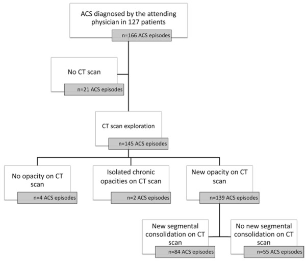
We screened 127 consecutive patients during 166 ACS episodes and 145 CT scans (in 118 consecutive patients) were included in the study.

RESULTS

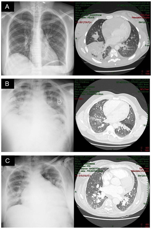
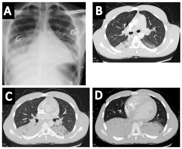
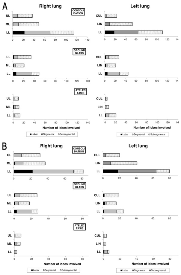
Among the 145 CT scans, 139 (96%) exhibited a new pulmonary opacity and 84 (58%) exhibited at least one complete lung segment consolidation. Consolidations were predominant as compared with ground-glass opacities and atelectasis. Lung parenchyma was increasingly consolidated from apex to base; the right and left inferior lobes were almost always involved in patients with a new complete lung segment consolidation on CT scan (98% and 95% of cases, respectively). Patients with a new complete lung segment consolidation on CT scan had a more severe presentation and course as compared with others. The sensitivity of bedside CR for the diagnosis of ACS using CT as a reference was good (>85%), whereas the specificity was weak (<60%).

CONCLUSIONS

ACS more frequently presented on CT as a consolidation pattern, predominating in lung bases. The reproducibility and diagnostic capacity of bedside CR were far from perfect. These findings may help improve the bedside imaging diagnosis of ACS.

摘要

介绍

镰状细胞病患者急性胸部综合征(ACS)的肺部计算机断层扫描(CT)特征尚未得到很好的描述,床边胸部 X 线摄影(CR)的诊断性能尚未得到检验。我们的目的是描述 ACS 的 CT 特征,并评估床边 CR 的可重复性和诊断性能。

方法

我们在 166 次 ACS 发作期间筛查了 127 例连续患者,共有 145 次 CT 扫描(118 例连续患者)纳入研究。

结果

在 145 次 CT 扫描中,139 次(96%)显示新的肺部不透明度,84 次(58%)显示至少一个完整肺段实变。实变比磨玻璃影和肺不张更为常见。肺实质从肺尖到肺底逐渐实变;在 CT 扫描上有新的完全肺段实变的患者中,右肺下叶和左肺下叶几乎总是受累(分别为 98%和 95%的病例)。与其他患者相比,在 CT 扫描上有新的完全肺段实变的患者表现更为严重,病程也更长。床边 CR 对 ACS 的诊断敏感性(以 CT 为参考)较好(>85%),但特异性较差(<60%)。

结论

ACS 在 CT 上更常表现为实变模式,以下肺为主。床边 CR 的可重复性和诊断能力远非完美。这些发现可能有助于改善 ACS 的床边影像学诊断。

https://cdn.ncbi.nlm.nih.gov/pmc/blobs/7fc0/5104836/20d9dcef54c1/halms950986f4.jpg
https://cdn.ncbi.nlm.nih.gov/pmc/blobs/7fc0/5104836/1a0a306eab59/halms950986f1.jpg
https://cdn.ncbi.nlm.nih.gov/pmc/blobs/7fc0/5104836/af172c1c994d/halms950986f2.jpg
https://cdn.ncbi.nlm.nih.gov/pmc/blobs/7fc0/5104836/762f4d960115/halms950986f3.jpg
https://cdn.ncbi.nlm.nih.gov/pmc/blobs/7fc0/5104836/20d9dcef54c1/halms950986f4.jpg