Nishio Jun, Ueki Takayuki, Naito Masatoshi
Department of Orthopaedic Surgery, Faculty of Medicine, Fukuoka University, Fukuoka 814-0180, Japan.
Exp Ther Med. 2013 Jul;6(1):164-166. doi: 10.3892/etm.2013.1084. Epub 2013 Apr 29.
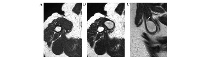
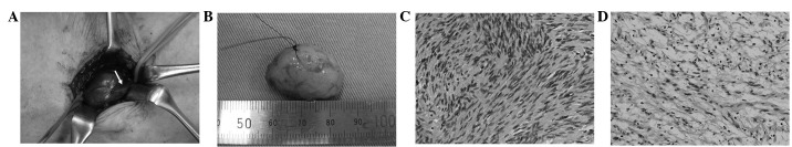
A schwannoma is a benign peripheral nerve sheath tumor composed exclusively of Schwann cells. A major-nerve schwannoma with an intramuscular location is an extremely rare condition. We present a rare case of intramuscular schwannoma originating from the musculocutaneous nerve in a 71-year-old female. The patient presented with a 7-month history of a slowly growing, painless mass in the medial aspect of the proximal upper arm. Magnetic resonance imaging revealed an oval-shaped intramuscular soft tissue mass with iso-signal intensity relative to skeletal muscle on T1-weighted images and high signal intensity on T2-weighted images. A rim of fat surrounding the mass, suggesting the split-fat sign, was also observed. The tumor was completely enucleated using an intracapsular technique. Histological examination confirmed the diagnosis of schwannoma consisting of Antoni A and B areas. There was no immediate neurological deficit following surgery. The patient had no evidence of local recurrence and no neurological deficit at final follow-up. To the best of our knowledge, this is the first report of musculocutaneous nerve schwannoma within the coracobrachialis muscle.
施万细胞瘤是一种仅由施万细胞组成的良性周围神经鞘瘤。肌内定位的主要神经施万细胞瘤是一种极其罕见的情况。我们报告一例71岁女性起源于肌皮神经的肌内施万细胞瘤的罕见病例。患者出现近端上臂内侧缓慢生长、无痛性肿块7个月病史。磁共振成像显示椭圆形肌内软组织肿块,在T1加权图像上相对于骨骼肌呈等信号强度,在T2加权图像上呈高信号强度。还观察到肿块周围有一圈脂肪,提示脂肪分裂征。采用囊内技术将肿瘤完整摘除。组织学检查确诊为施万细胞瘤,由Antoni A区和B区组成。术后无即刻神经功能缺损。在最后一次随访时,患者没有局部复发的证据,也没有神经功能缺损。据我们所知,这是肱二头肌内肌皮神经施万细胞瘤的首例报告。