Vafaie Faran, Yin Hao, O'Neil Caroline, Nong Zengxuan, Watson Alanna, Arpino John-Michael, Chu Michael W A, Wayne Holdsworth David, Gros Robert, Pickering J Geoffrey
Robarts Research Institute, Western University, London, ON, Canada; Departments of Medicine and Biochemistry, Western University, London, ON, Canada.
Aging Cell. 2014 Feb;13(1):121-30. doi: 10.1111/acel.12155. Epub 2013 Sep 19.
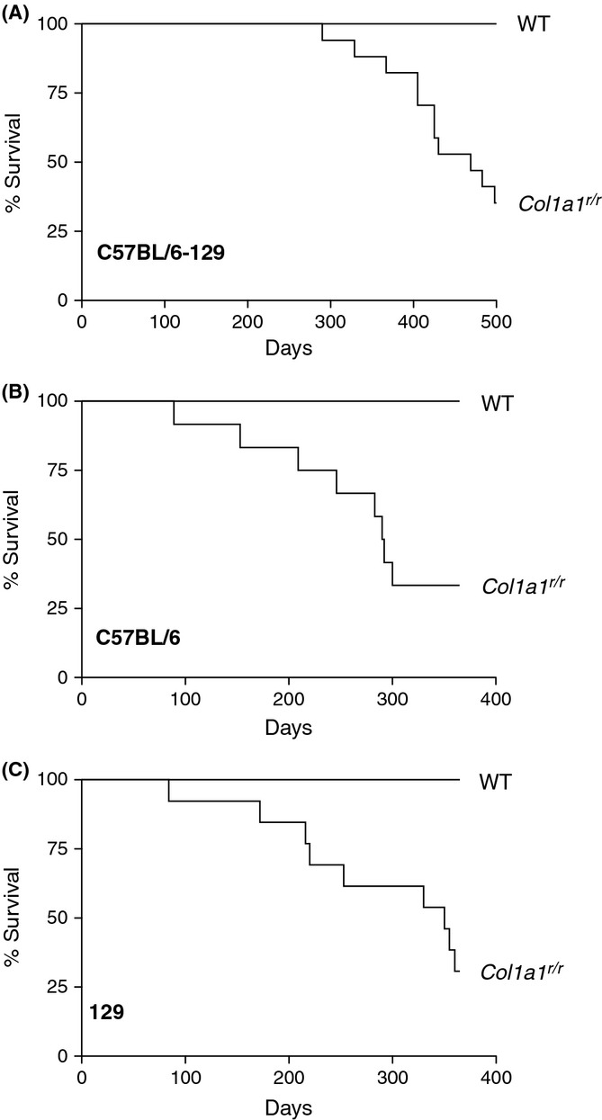
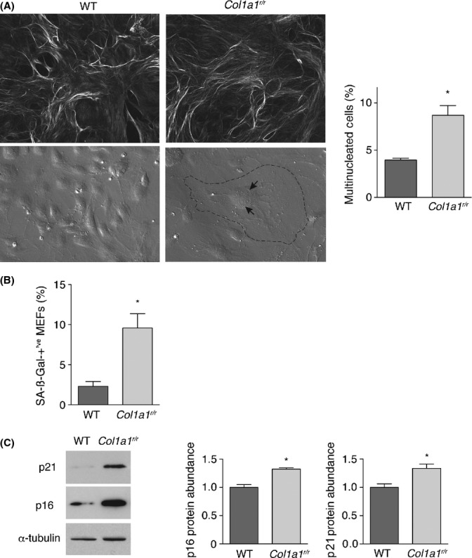
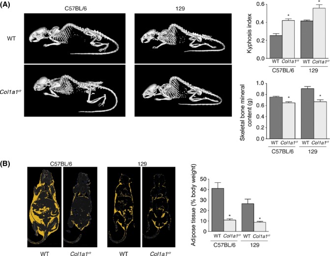
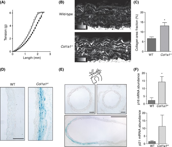
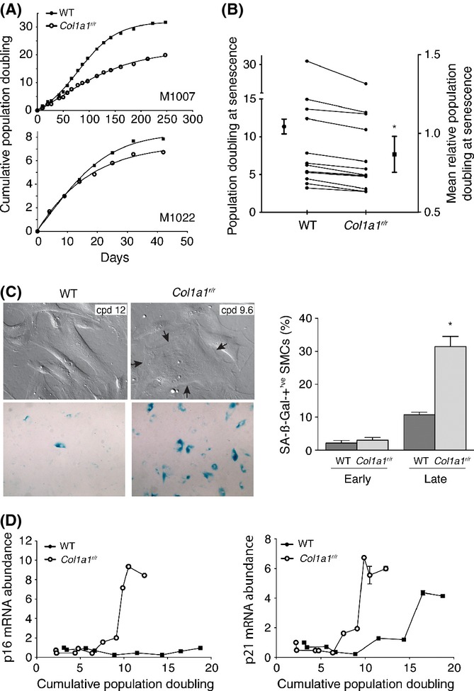
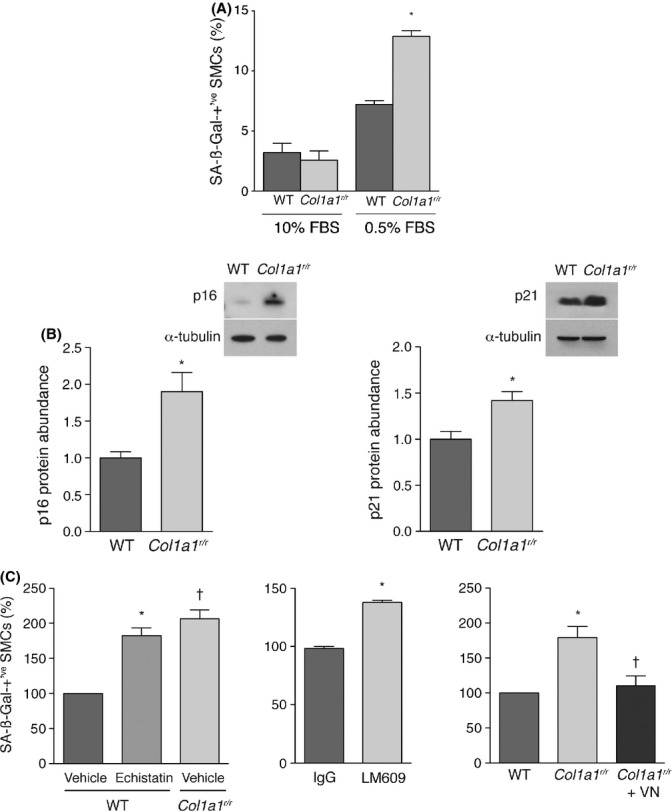
Collagen fibrils become resistant to cleavage over time. We hypothesized that resistance to type I collagen proteolysis not only marks biological aging but also drives it. To test this, we followed mice with a targeted mutation (Col1a1(r/r) ) that yields collagenase-resistant type I collagen. Compared with wild-type littermates, Col1a1(r/r) mice had a shortened lifespan and developed features of premature aging including kyphosis, weight loss, decreased bone mineral density, and hypertension. We also found that vascular smooth muscle cells (SMCs) in the aortic wall of Col1a1(r/r) mice were susceptible to stress-induced senescence, displaying senescence-associated ß-galactosidase (SA-ßGal) activity and upregulated p16(INK4A) in response to angiotensin II infusion. To elucidate the basis of this pro-aging effect, vascular SMCs from twelve patients undergoing coronary artery bypass surgery were cultured on collagen derived from Col1a1(r/r) or wild-type mice. This revealed that mutant collagen directly reduced replicative lifespan and increased stress-induced SA-ßGal activity, p16(INK4A) expression, and p21(CIP1) expression. The pro-senescence effect of mutant collagen was blocked by vitronectin, a ligand for αvß3 integrin that is presented by denatured but not native collagen. Moreover, inhibition of αvß3 with echistatin or with αvß3-blocking antibody increased senescence of SMCs on wild-type collagen. These findings reveal a novel aging cascade whereby resistance to collagen cleavage accelerates cellular aging. This interplay between extracellular and cellular compartments could hasten mammalian aging and the progression of aging-related diseases.
随着时间的推移,胶原纤维对裂解的抵抗力增强。我们推测,对I型胶原蛋白蛋白水解的抵抗力不仅标志着生物衰老,而且还推动着衰老进程。为了验证这一点,我们追踪了具有靶向突变(Col1a1(r/r))的小鼠,该突变产生抗胶原酶的I型胶原蛋白。与野生型同窝小鼠相比,Col1a1(r/r)小鼠的寿命缩短,并出现了早衰特征,包括脊柱后凸、体重减轻、骨矿物质密度降低和高血压。我们还发现,Col1a1(r/r)小鼠主动脉壁中的血管平滑肌细胞(SMC)易受应激诱导的衰老影响,在输注血管紧张素II后表现出衰老相关的β-半乳糖苷酶(SA-βGal)活性,并上调了p16(INK4A)。为了阐明这种促衰老作用的基础,将12例接受冠状动脉搭桥手术患者的血管SMC培养在源自Col1a1(r/r)或野生型小鼠的胶原蛋白上。结果显示,突变型胶原蛋白直接缩短了复制寿命,并增加了应激诱导的SA-βGal活性、p16(INK4A)表达和p21(CIP1)表达。玻连蛋白可阻断突变型胶原蛋白的促衰老作用,玻连蛋白是αvβ3整合素的配体,由变性而非天然胶原蛋白呈现。此外,用echistatin或αvβ3阻断抗体抑制αvβ3会增加野生型胶原蛋白上SMC的衰老。这些发现揭示了一种新的衰老级联反应,即对胶原蛋白裂解的抵抗力加速细胞衰老。细胞外和细胞内区室之间的这种相互作用可能会加速哺乳动物衰老和衰老相关疾病的进展。