Department of Internal Medicine & Yale Comprehensive Cancer Center, Yale University School of Medicine, New Haven, Connecticut, United States of America ; Medical Service, VA Connecticut Healthcare System, West Haven, Connecticut, United States of America.
PLoS One. 2013 Aug 19;8(8):e72032. doi: 10.1371/journal.pone.0072032. eCollection 2013.
Androgen deprivation therapy (ADT) is a common treatment for non-metastatic, low-risk prostate cancer, but a potential side effect of ADT is impaired brain functioning. Previous work with functional magnetic resonance imaging (MRI) demonstrated altered prefrontal cortical activations in cognitive control, with undetectable changes in behavioral performance. Given the utility of brain imaging in identifying the potentially deleterious effects of ADT on brain functions, the current study examined the effects of ADT on cerebral structures using high resolution MRI and voxel-based morphometry (VBM).
High resolution T1 weighted image of the whole brain were acquired at baseline and six months after ADT for 12 prostate cancer patients and 12 demographically matched non-exposed control participants imaged at the same time points. Brain images were segmented into gray matter, white matter and cerebral ventricles using the VBM toolbox as implemented in Statistical Parametric Mapping 8.
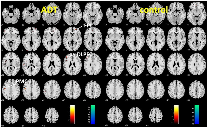
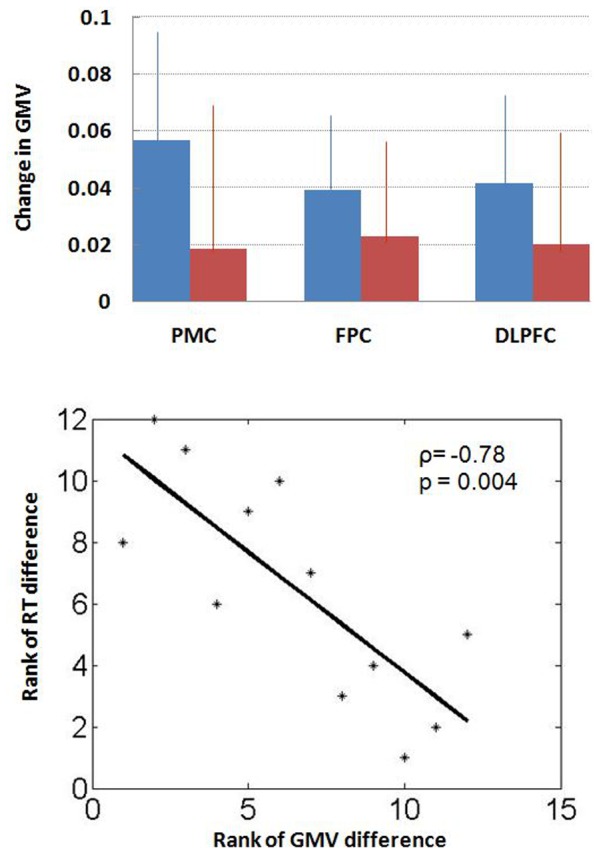
Compared to baseline scan, prostate cancer patients undergoing ADT showed decreased gray matter volume in frontopolar cortex, dorsolateral prefrontal cortex and primary motor cortex, whereas the non-exposed control participants did not show such changes. In addition, the decrease in gray matter volume of the primary motor cortex showed a significant correlation with longer reaction time to target detection in a working memory task.
ADT can affect cerebral gray matter volumes in prostate cancer patients. If replicated, these results may facilitate future studies of cognitive function and quality of life in men receiving ADT, and can also help clinicians weigh the benefits and risks of hormonal therapy in the treatment of prostate cancer.
雄激素剥夺疗法(ADT)是治疗非转移性、低危前列腺癌的常用方法,但 ADT 的一个潜在副作用是大脑功能受损。先前使用功能磁共振成像(fMRI)的研究表明,认知控制的前额叶皮质激活发生改变,而行为表现没有变化。鉴于脑成像在识别 ADT 对大脑功能的潜在有害影响方面的效用,本研究使用高分辨率磁共振成像和基于体素的形态测量学(VBM)来检查 ADT 对大脑结构的影响。
在 ADT 前和 ADT 后 6 个月,对 12 名前列腺癌患者和 12 名在相同时间点接受成像的年龄匹配的非暴露对照参与者采集全脑高分辨率 T1 加权图像。使用实施在统计参数映射 8 中的 VBM 工具包将脑图像分割为灰质、白质和脑室。
与基线扫描相比,接受 ADT 的前列腺癌患者在前额极皮质、背外侧前额叶皮质和初级运动皮质的灰质体积减少,而非暴露对照参与者则没有这种变化。此外,初级运动皮质的灰质体积减少与工作记忆任务中目标检测的反应时间延长呈显著相关。
ADT 可影响前列腺癌患者的大脑灰质体积。如果得到复制,这些结果可能有助于未来研究接受 ADT 的男性的认知功能和生活质量,并有助于临床医生权衡激素治疗在前列腺癌治疗中的益处和风险。