Tumor Virus Molecular Biology Section, Laboratory of Cellular Oncology, Center for Cancer Research, National Cancer Institute, National Institutes of Health, Bethesda, Maryland, United States of America.
PLoS Pathog. 2013;9(8):e1003558. doi: 10.1371/journal.ppat.1003558. Epub 2013 Aug 22.
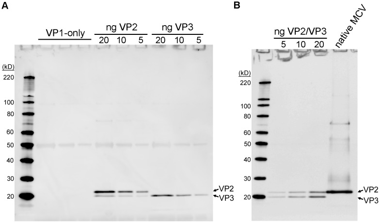
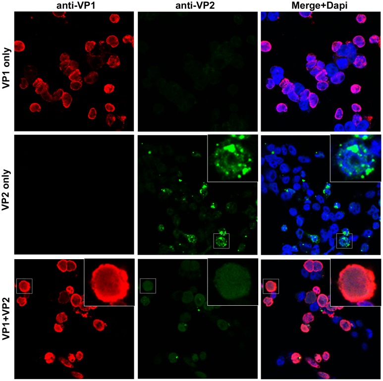
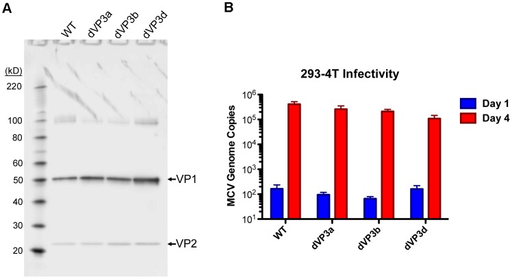
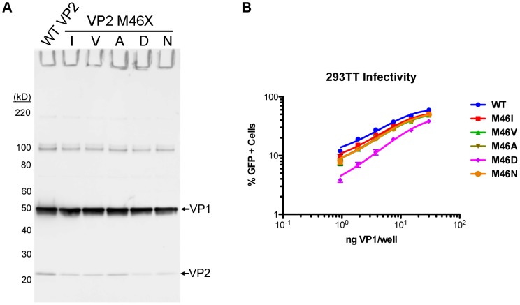
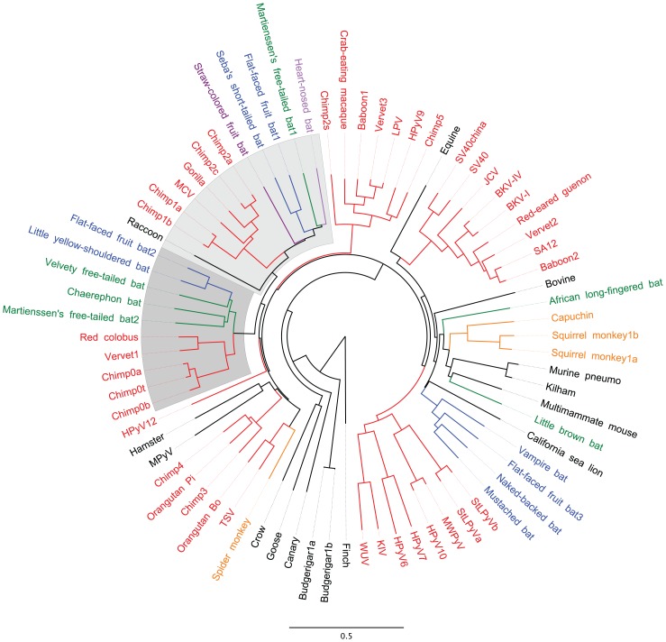
The surface of polyomavirus virions is composed of pentameric knobs of the major capsid protein, VP1. In previously studied polyomavirus species, such as SV40, two interior capsid proteins, VP2 and VP3, emerge from the virion to play important roles during the infectious entry process. Translation of the VP3 protein initiates at a highly conserved Met-Ala-Leu motif within the VP2 open reading frame. Phylogenetic analyses indicate that Merkel cell polyomavirus (MCV or MCPyV) is a member of a divergent clade of polyomaviruses that lack the conserved VP3 N-terminal motif. Consistent with this observation, we show that VP3 is not detectable in MCV-infected cells, VP3 is not found in native MCV virions, and mutation of possible alternative VP3-initiating methionine codons did not significantly affect MCV infectivity in culture. In contrast, VP2 knockout resulted in a >100-fold decrease in native MCV infectivity, despite normal virion assembly, viral DNA packaging, and cell attachment. Although pseudovirus-based experiments confirmed that VP2 plays an essential role for infection of some cell lines, other cell lines were readily transduced by pseudovirions lacking VP2. In cell lines where VP2 was needed for efficient infectious entry, the presence of a conserved myristoyl modification on the N-terminus of VP2 was important for its function. The results show that a single minor capsid protein, VP2, facilitates a post-attachment stage of MCV infectious entry into some, but not all, cell types.
多瘤病毒病毒体的表面由五聚体的主要衣壳蛋白 VP1 构成。在之前研究的多瘤病毒物种中,如 SV40,两种内部衣壳蛋白 VP2 和 VP3 从病毒体中出现,在感染进入过程中发挥重要作用。VP3 蛋白的翻译起始于 VP2 开放阅读框内高度保守的 Met-Ala-Leu 基序。系统发育分析表明,默克尔细胞多瘤病毒(MCV 或 MCPyV)是缺乏保守 VP3 N 端基序的多瘤病毒分化枝的成员。与这一观察结果一致,我们表明在 MCV 感染的细胞中检测不到 VP3,在天然 MCV 病毒体中也没有发现 VP3,并且突变可能的替代 VP3 起始甲硫氨酸密码子对 MCV 在培养物中的感染性没有显著影响。相比之下,尽管正常的病毒体组装、病毒 DNA 包装和细胞附着,VP2 缺失导致天然 MCV 感染力下降了 100 多倍。尽管如此,基于假病毒的实验证实 VP2 在某些细胞系的感染中起着至关重要的作用,但其他细胞系很容易被缺乏 VP2 的假病毒转导。在 VP2 对有效感染进入所必需的细胞系中,VP2 N 端保守的肉豆蔻酰化修饰对其功能很重要。结果表明,单一的次要衣壳蛋白 VP2 促进了 MCV 感染进入某些但不是所有细胞类型的附着后阶段。