Takeuchi Nobuhiro, Naba Kazuyoshi
Division of Gastroenterology, Department of Internal Medicine, Kawasaki Hospital, 3-3-1 Higashiyama-cho, Kobe, Hyogo 652-0042 Japan.
Clin J Gastroenterol. 2013 Aug;6(4):281-6. doi: 10.1007/s12328-013-0393-y. Epub 2013 Jun 13.
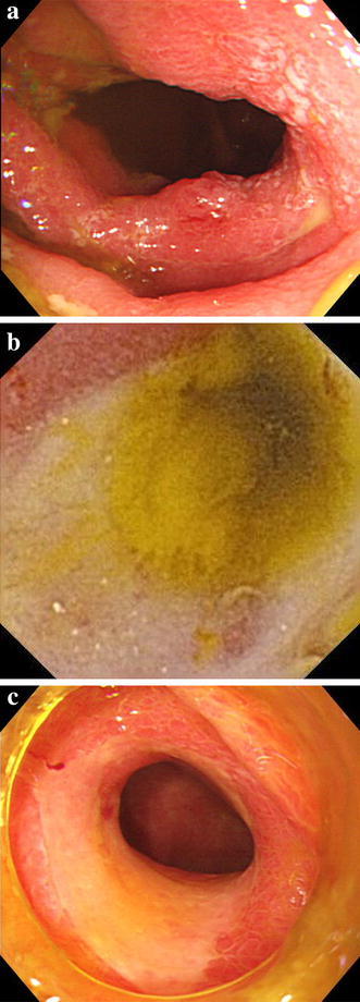
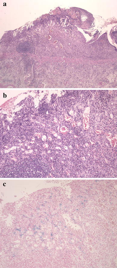
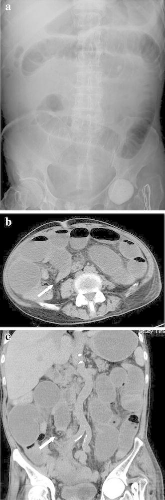
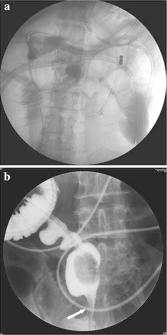
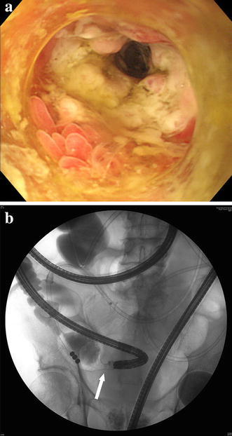
A 69-year-old male was admitted to our institution because of a sudden onset of vomiting and abdominal distention. His past history of illness included femoral head fracture, congestive heart failure and ischaemic colitis. Plain abdominal computed tomography revealed extensively dilated small intestinal loops with a calibre change around the end of the ileum. Small intestinal obstruction was diagnosed and a transnasal ileus tube was placed. The ileus tube was constantly moved towards small intestine until it reached the distal ileum. Contrast medium from the ileus tube revealed a distal ileal stricture. Subsequently, transanal single balloon enteroscopy was performed to inspect the stricture, revealing a circumferential and afferent tubular ulcer in the distal ileum, 5 cm from the ileocecal valve; gastrofluorography confirmed the stricture. Although the stricture was dilated on several occasions using balloon catheters, the stricture could not be improved. However, during the treatment, his general condition worsened over time; thus, surgical treatment was decided. Operative findings revealed several circumferential ulcers with a clear margin 5-28 cm from the ileocecal valve: all lesions were successfully resected. Pathological findings were consistent with ischaemic enteritis. We report a case of small intestinal obstruction resulting from stenotic ischaemic enteritis.
一名69岁男性因突然出现呕吐和腹胀入院。他既往有股骨头骨折、充血性心力衰竭和缺血性结肠炎病史。腹部平扫计算机断层扫描显示小肠肠袢广泛扩张,回肠末端周围肠腔有改变。诊断为小肠梗阻,并放置了经鼻肠梗阻导管。肠梗阻导管不断向小肠推进,直至到达回肠远端。来自肠梗阻导管的造影剂显示回肠远端狭窄。随后,进行经肛门单气囊小肠镜检查以检查狭窄部位,发现距回盲瓣5 cm处的回肠远端有一个环形和传入性管状溃疡;胃肠造影证实了狭窄。尽管使用球囊导管多次扩张狭窄部位,但狭窄仍无法改善。然而,在治疗过程中,他的一般状况随时间恶化;因此,决定进行手术治疗。手术发现距回盲瓣5 - 28 cm处有几个边缘清晰的环形溃疡:所有病变均成功切除。病理结果与缺血性肠炎一致。我们报告一例由狭窄性缺血性肠炎导致的小肠梗阻病例。