Zhou Yongxia, Lui Yvonne W, Zuo Xi-Nian, Milham Michael P, Reaume Joseph, Grossman Robert I, Ge Yulin
Department of Radiology / Center for Biomedical Imaging, NYU Langone Medical Center, New York, New York, USA.
J Magn Reson Imaging. 2014 Jun;39(6):1558-68. doi: 10.1002/jmri.24310. Epub 2013 Sep 6.
To examine thalamic and cortical injuries using fractional amplitude of low-frequency fluctuations (fALFFs) and functional connectivity MRI (fcMRI) based on resting state (RS) and task-related fMRI in patients with mild traumatic brain injury (MTBI).
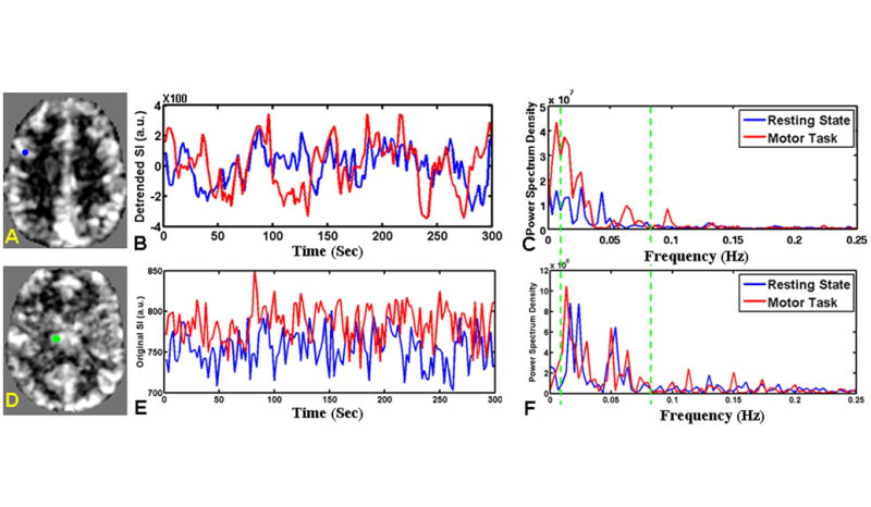
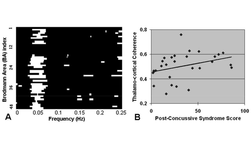
Twenty-seven patients and 27 age-matched controls were recruited. The 3 Tesla fMRI at RS and finger tapping task were used to assess fALFF and fcMRI patterns. fALFFs were computed with filtering (0.01-0.08 Hz) and scaling after preprocessing. fcMRI was performed using a standard seed-based correlation method, and delayed fcMRI (coherence) in frequency domain were also performed between thalamus and cortex.
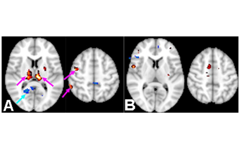
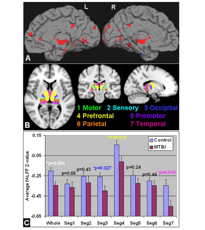
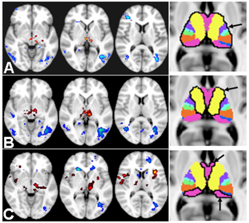
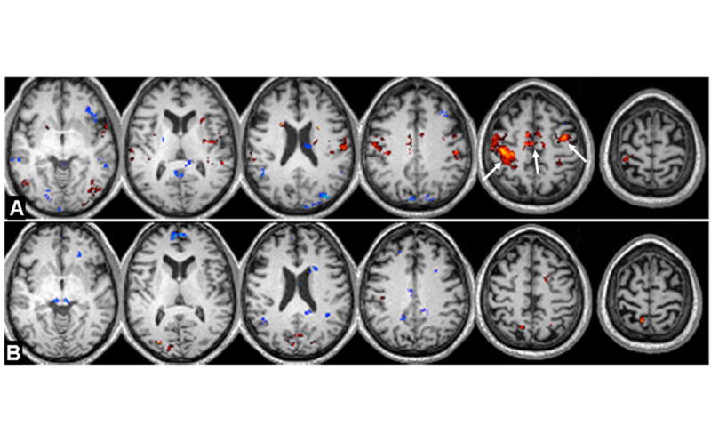
In comparison with controls, MTBI patients exhibited significantly decreased fALFFs in the thalamus (and frontal/temporal subsegments) and cortical frontal and temporal lobes; as well as decreased thalamo-thalamo and thalamo-frontal/ thalamo-temporal fcMRI at rest based on RS-fMRI (corrected P < 0.05). This thalamic and cortical disruption also existed at task-related condition in patients.
The decreased fALFFs (i.e., lower neuronal activity) in the thalamus and its segments provide additional evidence of thalamic injury in patients with MTBI. Our findings of fALFFs and fcMRI changes during motor task and resting state may offer insights into the underlying cause and primary location of disrupted thalamo-cortical networks after MTBI.
利用低频振幅分数(fALFF)和基于静息态(RS)及任务相关功能磁共振成像(fMRI)的功能连接磁共振成像(fcMRI),研究轻度创伤性脑损伤(MTBI)患者的丘脑和皮质损伤情况。
招募了27例患者和27例年龄匹配的对照者。使用3特斯拉静息态fMRI和手指敲击任务来评估fALFF和fcMRI模式。在预处理后,通过滤波(0.01 - 0.08赫兹)和缩放来计算fALFF。使用标准的基于种子点的相关方法进行fcMRI,并在丘脑和皮质之间进行频域延迟fcMRI(相干性)分析。
与对照组相比,MTBI患者丘脑(以及额叶/颞叶亚段)、皮质额叶和颞叶的fALFF显著降低;基于静息态fMRI的静息状态下,丘脑-丘脑、丘脑-额叶/丘脑-颞叶的fcMRI也降低(校正P < 0.05)。这种丘脑和皮质的破坏在患者的任务相关状态下也存在。
丘脑及其亚段fALFF降低(即神经元活动较低)为MTBI患者的丘脑损伤提供了额外证据。我们关于运动任务和静息状态期间fALFF和fcMRI变化的研究结果,可能有助于深入了解MTBI后丘脑-皮质网络破坏的潜在原因和主要部位。