1] Department of Microbiology and Immunology, Nippon Medical School, Tokyo, Japan [2] Third Department of Internal Medicine, Nippon Medical School, Tokyo, Japan.
Immunol Cell Biol. 2013 Oct;91(9):545-55. doi: 10.1038/icb.2013.38. Epub 2013 Sep 10.
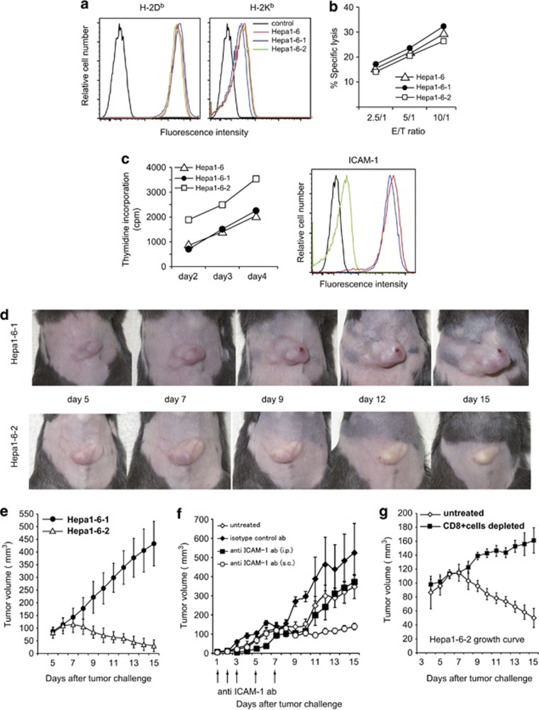
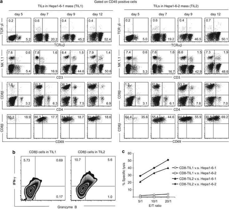
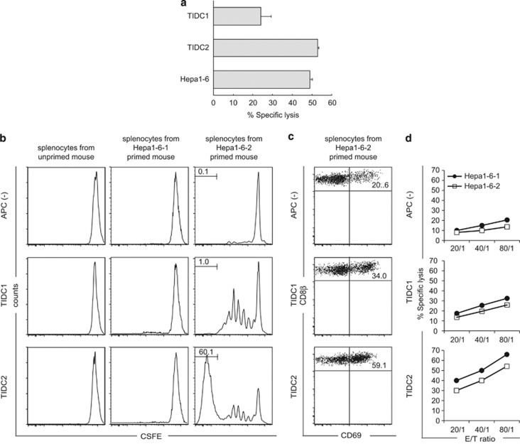
Cancer immunosurveillance failure is largely attributed to the insufficient activation of tumor-specific class I major histocompatibility complex (MHC) molecule (MHC-I)-restricted CD8⁺ cytotoxic T lymphocytes (CTLs). DEC-205⁺ dendritic cells (DCs), having the ability to cross-present, can present captured tumor antigens on MHC-I alongside costimulatory molecules, inducing the priming and activation of tumor-specific CD8⁺ CTLs. It has been suggested that reduced levels of costimulatory molecules on DCs may be a cause of impaired CTL induction and that some tumors may induce the downregulation of costimulatory molecules on tolerogenic DCs. To examine such possibilities, we established two distinct types of murine hepatoma cell lines, named Hepa1-6-1 and Hepa1-6-2 (derived from Hepa1-6 cells), and confirmed that they display similar antigenicities, as well as identical surface expression of MHC-I. We found that Hepa1-6-1 had the ability to grow continuously after subcutaneous implantation into syngeneic C57BL/6 mice and did not prime CD8⁺ CTLs. In contrast, Hepa1-6-2 cells, which display reduced levels of adhesion molecules, such as Intercellular Adhesion Molecule 1 (ICAM-1), failed to grow in vivo and efficiently primed CTLs. Moreover, Hepa1-6-1-derived factors, such as transforming growth factor (TGF)-β1, vascular endothelial growth factor (VEGF) and α-fetoprotein (AFP), converted CD11c(high) MHC-II(high) DEC-205⁺ DC subsets into tolerogenic cells, displaying downregulated costimulatory molecules and having impaired cross-presenting capacities. These immunosuppressive tolerogenic DCs appeared to inhibit the induction of tumor-specific CD8⁺ CTLs and suppress their cytotoxic functions within the tumor. Together, the findings presented here provide a new method of cancer immunotherapy using the selective suppression, depletion or alteration of immunosuppressive tolerogenic DCs within tumors.
肿瘤免疫监视失败在很大程度上归因于肿瘤特异性 I 类主要组织相容性复合体 (MHC) 分子 (MHC-I) 受限的 CD8⁺细胞毒性 T 淋巴细胞 (CTL) 激活不足。具有交叉呈递能力的 DEC-205⁺树突状细胞 (DC) 可以在 MHC-I 上与共刺激分子一起呈递捕获的肿瘤抗原,诱导肿瘤特异性 CD8⁺CTL 的启动和激活。有人提出,DC 上共刺激分子水平降低可能是 CTL 诱导受损的原因,并且一些肿瘤可能诱导耐受型 DC 上共刺激分子的下调。为了检查这种可能性,我们建立了两种不同类型的小鼠肝癌细胞系,命名为 Hepa1-6-1 和 Hepa1-6-2(源自 Hepa1-6 细胞),并证实它们具有相似的抗原性,以及相同的 MHC-I 表面表达。我们发现 Hepa1-6-1 具有在皮下植入同基因 C57BL/6 小鼠后连续生长的能力,并且不能诱导 CD8⁺CTL。相比之下,Hepa1-6-2 细胞,其黏附分子(如细胞间黏附分子 1(ICAM-1))水平降低,在体内无法生长并有效诱导 CTL。此外,Hepa1-6-1 衍生的因子,如转化生长因子 (TGF)-β1、血管内皮生长因子 (VEGF) 和甲胎蛋白 (AFP),将 CD11c(高)MHC-II(高)DEC-205⁺DC 亚群转化为耐受型细胞,表现出下调的共刺激分子和受损的交叉呈递能力。这些免疫抑制性耐受型 DC 似乎抑制了肿瘤特异性 CD8⁺CTL 的诱导,并抑制了它们在肿瘤内的细胞毒性功能。总之,这里提出的发现为使用选择性抑制、耗尽或改变肿瘤内免疫抑制性耐受型 DC 提供了一种新的癌症免疫疗法。