• 文献检索
  • 文档翻译
  • 深度研究
  • 学术资讯
  • Suppr Zotero 插件Zotero 插件
  • 邀请有礼
  • 套餐&价格
  • 历史记录
应用&插件
Suppr Zotero 插件Zotero 插件浏览器插件Mac 客户端Windows 客户端微信小程序
定价
高级版会员购买积分包购买API积分包
服务
文献检索文档翻译深度研究API 文档MCP 服务
关于我们
关于 Suppr公司介绍联系我们用户协议隐私条款
关注我们

Suppr 超能文献

核心技术专利:CN118964589B侵权必究
粤ICP备2023148730 号-1Suppr @ 2026

文献检索

告别复杂PubMed语法,用中文像聊天一样搜索,搜遍4000万医学文献。AI智能推荐,让科研检索更轻松。

立即免费搜索

文件翻译

保留排版,准确专业,支持PDF/Word/PPT等文件格式,支持 12+语言互译。

免费翻译文档

深度研究

AI帮你快速写综述,25分钟生成高质量综述,智能提取关键信息,辅助科研写作。

立即免费体验

单次支气管镜检查过程中获取的二次冲洗标本在肺结核诊断中的额外作用。

Additional role of second washing specimen obtained during single bronchoscopy session in diagnosis of pulmonary tuberculosis.

作者信息

Yoo Hongseok, Song Jae-Uk, Koh Won-Jung, Jeon Kyeongman, Um Sang-Won, Suh Gee Young, Chung Man Pyo, Kim Hojoong, Kwon O Jung, Lee Nam Yong, Woo Sookyoung, Park Hye Yun

机构信息

Division of Pulmonary and Critical Care Medicine, Department of Medicine, Samsung Medical Center, Sungkyunkwan University School of Medicine, 81 Irwon-ro, Gangnam-gu, Seoul, Republic of Korea.

出版信息

BMC Infect Dis. 2013 Sep 2;13:404. doi: 10.1186/1471-2334-13-404.

DOI:10.1186/1471-2334-13-404
PMID:24059248
原文链接:https://pmc.ncbi.nlm.nih.gov/articles/PMC3765986/
Abstract

BACKGROUND

Flexible bronchoscopy with bronchial washing is a useful procedure for diagnosis of pulmonary tuberculosis (TB), when a patient cannot produce sputum spontaneously or when sputum smears are negative. However, the benefit of gaining serial bronchial washing specimens for diagnosis of TB has not yet been studied. Therefore, we conducted a retrospective study to determine the diagnostic utility of additional bronchial washing specimens for the diagnosis of pulmonary TB in suspected patients.

METHODS

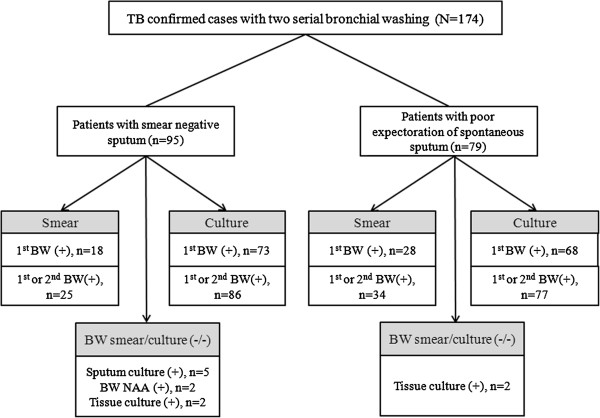
A retrospective analysis was performed on 174 patients [sputum smear-negative, n = 95 (55%); lack of sputum specimen, n = 79 (45%)] who received flexible bronchoscopy with two bronchial washing specimens with microbiological confirmation of pulmonary TB in Samsung Medical Center, between January, 2010 and December, 2011.

RESULTS

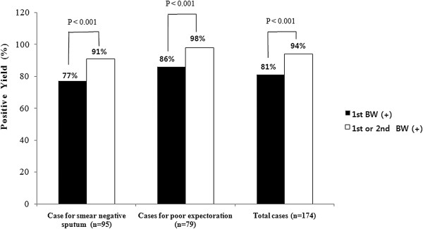
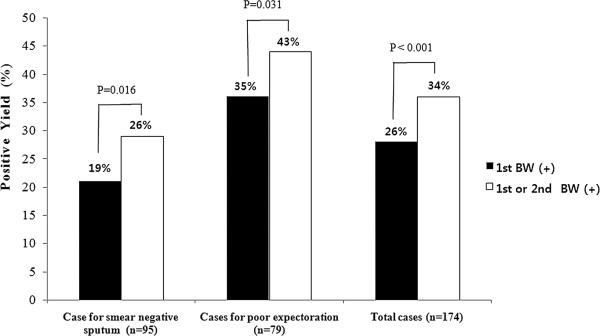
Pulmonary TB was diagnosed by first bronchial washing specimen in 141 patients (81%) out of 174 enrolled patients, and an additional bronchial washing specimen established diagnosis exclusively in 22 (13%) patients. Smear for acid-fast bacilli (AFB) was positive in 46 patients (26%) for the first bronchial washing specimen. Thirteen patients (7%) were positive only on smear of an additional bronchial washing specimen. Combined smear positivity of the first and second bronchial washing specimens was significantly higher compared to first bronchial washing specimen alone [Total cases: 59 (34%) vs. 46 (26%), p < 0.001; cases for smear negative sputum: 25 (26%) vs. 18 (19%), p = 0.016; cases for poor expectoration: 34 (43%) vs. 28 (35%), p = 0.031]. The diagnostic yield determined by culture was also significantly higher in combination of the first and second bronchial washing specimens compared to the first bronchial washing. [Total cases: 163 (94%) vs. 141 (81%), p < 0.001; cases for smear negative sputum: 86 (91%) vs. 73 (77%), p < 0.001; cases for poor expectoration: 77 (98%) vs. 68 (86%), p = 0.004].

CONCLUSIONS

Obtaining an additional bronchial washing specimen could be a beneficial and considerable option for diagnosis of TB in patients with smear-negative sputum or who cannot produce sputum samples.

摘要

背景

当患者无法自行咳痰或痰涂片为阴性时,可弯曲支气管镜检查及支气管灌洗术是诊断肺结核(TB)的一种有用方法。然而,获取系列支气管灌洗标本对肺结核诊断的益处尚未得到研究。因此,我们进行了一项回顾性研究,以确定在疑似患者中额外的支气管灌洗标本对肺结核诊断的效用。

方法

对2010年1月至2011年12月期间在三星医疗中心接受可弯曲支气管镜检查并获取两份支气管灌洗标本且经微生物学确诊为肺结核的174例患者[痰涂片阴性,n = 95例(55%);无痰标本,n = 79例(45%)]进行回顾性分析。

结果

在174例纳入患者中,141例(81%)通过首次支气管灌洗标本确诊为肺结核,另外22例(13%)仅通过额外的支气管灌洗标本确诊。首次支气管灌洗标本中46例(26%)抗酸杆菌(AFB)涂片阳性。13例(7%)仅在额外支气管灌洗标本涂片上呈阳性。与仅首次支气管灌洗标本相比,首次和第二次支气管灌洗标本的联合涂片阳性率显著更高[总病例数:59例(34%)对46例(26%),p < 0.001;痰涂片阴性病例:25例(26%)对18例(19%),p = 0.016;咳痰不佳病例:34例(43%)对28例(35%),p = 0.031]。与首次支气管灌洗相比,首次和第二次支气管灌洗标本联合培养确定的诊断率也显著更高。[总病例数:163例(94%)对141例(81%),p < 0.001;痰涂片阴性病例:86例(91%)对73例(77%),p < 0.001;咳痰不佳病例:77例(98%)对68例(86%),p = 0.004]。

结论

对于痰涂片阴性或无法咳出痰标本的患者,获取额外的支气管灌洗标本可能是诊断肺结核的一种有益且重要的选择。

https://cdn.ncbi.nlm.nih.gov/pmc/blobs/2bad/3765986/fd9d204cbddc/1471-2334-13-404-3.jpg
https://cdn.ncbi.nlm.nih.gov/pmc/blobs/2bad/3765986/f7d8c12b792c/1471-2334-13-404-1.jpg
https://cdn.ncbi.nlm.nih.gov/pmc/blobs/2bad/3765986/b9a323623910/1471-2334-13-404-2.jpg
https://cdn.ncbi.nlm.nih.gov/pmc/blobs/2bad/3765986/fd9d204cbddc/1471-2334-13-404-3.jpg
https://cdn.ncbi.nlm.nih.gov/pmc/blobs/2bad/3765986/f7d8c12b792c/1471-2334-13-404-1.jpg
https://cdn.ncbi.nlm.nih.gov/pmc/blobs/2bad/3765986/b9a323623910/1471-2334-13-404-2.jpg
https://cdn.ncbi.nlm.nih.gov/pmc/blobs/2bad/3765986/fd9d204cbddc/1471-2334-13-404-3.jpg

相似文献

1
Additional role of second washing specimen obtained during single bronchoscopy session in diagnosis of pulmonary tuberculosis.单次支气管镜检查过程中获取的二次冲洗标本在肺结核诊断中的额外作用。
BMC Infect Dis. 2013 Sep 2;13:404. doi: 10.1186/1471-2334-13-404.
2
Incremental yield of bronchial washing for diagnosing smear-negative pulmonary tuberculosis.支气管灌洗对诊断菌阴肺结核的增量收益。
Rev Saude Publica. 2013 Aug;47(4):813-6. doi: 10.1590/S0034-8910.2013047004548.
3
Diagnostic value of bronchoalveolar lavage and bronchial washing in sputum-scarce or smear-negative cases with suspected pulmonary tuberculosis: a randomized study.疑似肺结核患者痰少或涂片阴性时支气管肺泡灌洗和支气管冲洗的诊断价值:一项随机研究。
Clin Microbiol Infect. 2020 Jul;26(7):911-916. doi: 10.1016/j.cmi.2019.11.013. Epub 2019 Nov 20.
4
Diagnostic yield of post-bronchoscopy sputum smear in pulmonary tuberculosis.支气管镜检查后痰涂片在肺结核诊断中的阳性率
Scand J Infect Dis. 2012 May;44(5):369-73. doi: 10.3109/00365548.2011.643820.
5
Usefulness of Mycobacterium tuberculosis-polymerase chain reaction with bronchial washing samples in predicting discontinuation of airborne infection isolation in patients hospitalized with suspected pulmonary tuberculosis.支气管灌洗液结核分枝杆菌聚合酶链反应在预测疑似肺结核住院患者空气传播感染隔离停止中的作用。
PLoS One. 2022 Dec 30;17(12):e0279256. doi: 10.1371/journal.pone.0279256. eCollection 2022.
6
Diagnosing sputum/smear-negative pulmonary tuberculosis: Does fibre-optic bronchoscopy play a significant role?诊断痰涂片阴性肺结核:纤维支气管镜检查是否发挥重要作用?
Lung India. 2010 Apr;27(2):58-62. doi: 10.4103/0970-2113.63607.
7
Usefulness of induced sputum and fibreoptic bronchoscopy specimens in the diagnosis of pulmonary tuberculosis.诱导痰和纤维支气管镜检查标本在肺结核诊断中的应用价值。
J Int Med Res. 2005 Mar-Apr;33(2):260-5. doi: 10.1177/147323000503300215.
8
The value of fiberoptic bronchoscopy in culture-positive pulmonary tuberculosis patients whose pre-bronchoscopic sputum specimens were negative both for smear and PCR analyses.纤维支气管镜检查对于支气管镜检查前痰标本涂片和PCR分析均为阴性的培养阳性肺结核患者的价值。
Intern Med. 2010;49(2):95-102. doi: 10.2169/internalmedicine.49.2686. Epub 2010 Jan 15.
9
Diagnostic accuracy of Xpert® MTB/RIF on bronchoscopy specimens in patients with suspected pulmonary tuberculosis.Xpert® MTB/RIF 检测对疑似肺结核患者支气管镜标本的诊断准确性。
Int J Tuberc Lung Dis. 2013 Jul;17(7):917-21. doi: 10.5588/ijtld.12.0885. Epub 2013 Apr 23.
10
[The value of fiberoptic bronchoscopy in diagnosis of smear negative pulmonary tuberculosis].[纤维支气管镜检查在痰涂片阴性肺结核诊断中的价值]
Tuberk Toraks. 2003;51(4):405-9.

引用本文的文献

1
Bronchoscopic Strategies to Improve Diagnostic Yield in Pulmonary Tuberculosis Patients.提高肺结核患者诊断率的支气管镜检查策略
Tuberc Respir Dis (Seoul). 2024 Jul;87(3):302-308. doi: 10.4046/trd.2024.0020. Epub 2024 Mar 28.
2
Additional Usefulness of Bronchoscopy in Patients with Initial Microbiologically Negative Pulmonary Tuberculosis: A Retrospective Analysis of a Korean Nationwide Prospective Cohort Study.支气管镜检查在初治微生物学阴性肺结核患者中的额外效用:一项韩国全国前瞻性队列研究的回顾性分析
Infect Drug Resist. 2022 Mar 12;15:1029-1037. doi: 10.2147/IDR.S354962. eCollection 2022.
3
Application of endobronchial ultrasonography using a guide sheath and electromagnetic navigation bronchoscopy in the diagnosis of atypical bacteriologically-negative pulmonary tuberculosis.

本文引用的文献

1
Fiberoptic bronchoscopy for the rapid diagnosis of smear-negative pulmonary tuberculosis.纤维支气管镜检查对菌阴肺结核的快速诊断。
BMC Infect Dis. 2012 Jun 22;12:141. doi: 10.1186/1471-2334-12-141.
2
Incremental yield in sputum smear positivity by examining a second early morning sputum specimen in follow-up patients on DOTS: 7 year analysis of RNTCP laboratory register.在直接观察短程治疗(DOTS)随访患者中,通过检查第二份清晨痰标本提高痰涂片阳性率:对印度国家结核病控制规划(RNTCP)实验室登记册的7年分析
Indian J Tuberc. 2011 Apr;58(2):60-5.
3
Diagnosing sputum/smear-negative pulmonary tuberculosis: Does fibre-optic bronchoscopy play a significant role?
使用引导鞘的支气管内超声检查和电磁导航支气管镜检查在非典型细菌学阴性肺结核诊断中的应用
Ann Transl Med. 2019 Oct;7(20):567. doi: 10.21037/atm.2019.09.37.
4
Exposure to Mycobacterium tuberculosis during Flexible Bronchoscopy in Patients with Unexpected Pulmonary Tuberculosis.意外肺结核患者在柔性支气管镜检查期间暴露于结核分枝杆菌的情况。
PLoS One. 2016 May 26;11(5):e0156385. doi: 10.1371/journal.pone.0156385. eCollection 2016.
诊断痰涂片阴性肺结核:纤维支气管镜检查是否发挥重要作用?
Lung India. 2010 Apr;27(2):58-62. doi: 10.4103/0970-2113.63607.
4
Performance of nucleic acid amplification tests for diagnosis of tuberculosis in a large urban setting.大型城市环境中核酸扩增试验在结核病诊断中的性能
Clin Infect Dis. 2009 Jul 1;49(1):46-54. doi: 10.1086/599037.
5
Fibreoptic bronchoscopy in the diagnosis of sputum smear-negative pulmonary tuberculosis: current status.纤维支气管镜在痰涂片阴性肺结核诊断中的现状
Indian J Chest Dis Allied Sci. 2008 Jan-Mar;50(1):67-78.
6
The value of routinely culturing for tuberculosis during bronchoscopies in an intermediate tuberculosis-burden country.在一个结核病负担中等的国家,支气管镜检查期间常规进行结核培养的价值。
Yonsei Med J. 2007 Dec 31;48(6):969-72. doi: 10.3349/ymj.2007.48.6.969.
7
A method to determine the utility of the third diagnostic and the second follow-up sputum smear examinations to diagnose tuberculosis cases and failures.一种确定第三次诊断性和第二次随访痰涂片检查在诊断结核病例及治疗失败情况中的效用的方法。
Int J Tuberc Lung Dis. 2005 Apr;9(4):384-91.
8
Yield of smear, culture and amplification tests from repeated sputum induction for the diagnosis of pulmonary tuberculosis.重复痰液诱导进行涂片、培养及扩增检测对肺结核诊断的阳性率
Int J Tuberc Lung Dis. 2001 Sep;5(9):855-60.
9
Diagnostic yield of repeated smear microscopy examinations among patients suspected of pulmonary TB in Shandong province of China.中国山东省疑似肺结核患者重复涂片显微镜检查的诊断率
Int J Tuberc Lung Dis. 2000 Nov;4(11):1086-7.
10
Screening tuberculosis suspects using two sputum smears.使用两份痰涂片筛查结核病疑似患者。
Int J Tuberc Lung Dis. 2000 Jan;4(1):36-40.