• 文献检索
  • 文档翻译
  • 深度研究
  • 学术资讯
  • Suppr Zotero 插件Zotero 插件
  • 邀请有礼
  • 套餐&价格
  • 历史记录
应用&插件
Suppr Zotero 插件Zotero 插件浏览器插件Mac 客户端Windows 客户端微信小程序
定价
高级版会员购买积分包购买API积分包
服务
文献检索文档翻译深度研究API 文档MCP 服务
关于我们
关于 Suppr公司介绍联系我们用户协议隐私条款
关注我们

Suppr 超能文献

核心技术专利:CN118964589B侵权必究
粤ICP备2023148730 号-1Suppr @ 2026

文献检索

告别复杂PubMed语法,用中文像聊天一样搜索,搜遍4000万医学文献。AI智能推荐,让科研检索更轻松。

立即免费搜索

文件翻译

保留排版,准确专业,支持PDF/Word/PPT等文件格式,支持 12+语言互译。

免费翻译文档

深度研究

AI帮你快速写综述,25分钟生成高质量综述,智能提取关键信息,辅助科研写作。

立即免费体验

应用电影心血管磁共振评估分数面积变化率评价左心室舒张功能:一项可行性研究。

Evaluation of left ventricular diastolic function by fractional area change using cine cardiovascular magnetic resonance: a feasibility study.

机构信息

First Department of Internal Medicine, Nara Medical University, Nara, Japan.

出版信息

J Cardiovasc Magn Reson. 2013 Sep 26;15(1):87. doi: 10.1186/1532-429X-15-87.

DOI:10.1186/1532-429X-15-87
PMID:24070403
原文链接:https://pmc.ncbi.nlm.nih.gov/articles/PMC3815234/
Abstract

BACKGROUND

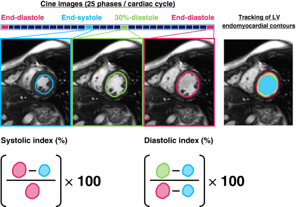
Evaluation of left ventricular (LV) diastolic function is essential for the management of heart failure. We verified whether LV diastolic function could be evaluated by measuring the fractional area change (FAC) using cine cardiovascular magnetic resonance (CMR).

METHODS

We collected clinical data from 59 patients who underwent echocardiography and cine CMR. Normal, impaired relaxation, pseudonormal, and restrictive LV filling were observed in 15, 28, 11, and 5 patients, respectively. We calculated FAC during the first 30% of diastole (diastolic-index%) in the short-axis view, by tracing the contours on only three MR cine images.

RESULTS

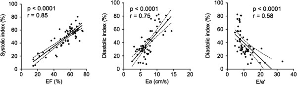
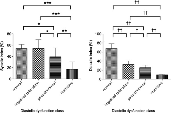
The diastolic index was significantly lower (p < 0.0001) in patients with impaired relaxation (32.4 ± 7.5), pseudonormal filling (25.4 ± 5.6), and restrictive filling (9.5 ± 1.5) compared to those with normal diastolic function (67.7 ± 10.8), and the index decreased significantly with worsening of diastolic dysfunction. The diastolic index correlated positively with early diastolic mitral annular velocity measured by tissue Doppler imaging (r = 0.75, p < 0.0001), respectively.

CONCLUSIONS

Measurement of FAC can be useful for the evaluation of LV diastolic function using cine CMR.

摘要

背景

评估左心室(LV)舒张功能对于心力衰竭的管理至关重要。我们通过测量电影心血管磁共振(CMR)的分数面积变化(FAC)来验证是否可以评估 LV 舒张功能。

方法

我们收集了 59 例接受超声心动图和电影 CMR 的患者的临床数据。在 15 例、28 例、11 例和 5 例患者中分别观察到正常、松弛不良、假性正常和限制性 LV 充盈。我们通过仅追踪三个 MR 电影图像上的轮廓,计算短轴视图中舒张期前 30%(舒张指数%)的 FAC。

结果

与舒张功能正常的患者(67.7 ± 10.8)相比,松弛不良(32.4 ± 7.5)、假性正常充盈(25.4 ± 5.6)和限制性充盈(9.5 ± 1.5)患者的舒张指数显著降低(p < 0.0001),舒张功能障碍程度越严重,舒张指数越低。舒张指数与组织多普勒成像测量的早期舒张二尖瓣环速度呈正相关(r = 0.75,p < 0.0001)。

结论

使用电影 CMR 测量 FAC 可用于评估 LV 舒张功能。

https://cdn.ncbi.nlm.nih.gov/pmc/blobs/61e9/3815234/ad4552224fbb/1532-429X-15-87-3.jpg
https://cdn.ncbi.nlm.nih.gov/pmc/blobs/61e9/3815234/d469518fc852/1532-429X-15-87-1.jpg
https://cdn.ncbi.nlm.nih.gov/pmc/blobs/61e9/3815234/b2d34e2528b9/1532-429X-15-87-2.jpg
https://cdn.ncbi.nlm.nih.gov/pmc/blobs/61e9/3815234/ad4552224fbb/1532-429X-15-87-3.jpg
https://cdn.ncbi.nlm.nih.gov/pmc/blobs/61e9/3815234/d469518fc852/1532-429X-15-87-1.jpg
https://cdn.ncbi.nlm.nih.gov/pmc/blobs/61e9/3815234/b2d34e2528b9/1532-429X-15-87-2.jpg
https://cdn.ncbi.nlm.nih.gov/pmc/blobs/61e9/3815234/ad4552224fbb/1532-429X-15-87-3.jpg

相似文献

1
Evaluation of left ventricular diastolic function by fractional area change using cine cardiovascular magnetic resonance: a feasibility study.应用电影心血管磁共振评估分数面积变化率评价左心室舒张功能:一项可行性研究。
J Cardiovasc Magn Reson. 2013 Sep 26;15(1):87. doi: 10.1186/1532-429X-15-87.
2
Evaluation of diastolic function by three-dimensional volume tracking of the mitral annulus with cardiovascular magnetic resonance: comparison with tissue Doppler imaging.利用心血管磁共振成像通过二尖瓣环三维容积追踪评估舒张功能:与组织多普勒成像的比较
J Cardiovasc Magn Reson. 2014 Sep 20;16(1):71. doi: 10.1186/s12968-014-0071-3.
3
Impact of diastolic dysfunction severity on global left ventricular volumetric filling - assessment by automated segmentation of routine cine cardiovascular magnetic resonance.常规电影心血管磁共振自动分割评估舒张功能障碍严重程度对整体左心室容积充盈的影响。
J Cardiovasc Magn Reson. 2010 Jul 31;12(1):46. doi: 10.1186/1532-429X-12-46.
4
Differentiation of left ventricular diastolic dysfunction, identification of pseudonormal/restrictive mitral inflow pattern and determination of left ventricular filling pressure by Tei index obtained from tissue Doppler echocardiography.左心室舒张功能障碍的鉴别、假性正常/限制性二尖瓣血流模式的识别以及通过组织多普勒超声心动图获得的Tei指数测定左心室充盈压。
Echocardiography. 2006 Apr;23(4):287-94. doi: 10.1111/j.1540-8175.2006.00222.x.
5
Comparison of cine magnetic resonance imaging and Doppler echocardiography for evaluation of left ventricular diastolic function.
Am J Cardiol. 1997 Aug 1;80(3):384-6. doi: 10.1016/s0002-9149(97)00375-5.
6
Early detection of subclinical ventricular deterioration in aortic stenosis with cardiovascular magnetic resonance and echocardiography.采用心血管磁共振和超声心动图早期检测主动脉瓣狭窄的亚临床心室恶化。
J Cardiovasc Magn Reson. 2013 Aug 28;15(1):72. doi: 10.1186/1532-429X-15-72.
7
Clinical aspects of left ventricular diastolic function assessed by Doppler echocardiography following acute myocardial infarction.急性心肌梗死后经多普勒超声心动图评估左心室舒张功能的临床方面
Dan Med Bull. 2001 Nov;48(4):199-210.
8
Automated left ventricular diastolic function evaluation from phase-contrast cardiovascular magnetic resonance and comparison with Doppler echocardiography.基于相位对比心血管磁共振的左心室舒张功能自动评估,并与多普勒超声心动图比较。
J Cardiovasc Magn Reson. 2010 Nov 9;12(1):63. doi: 10.1186/1532-429X-12-63.
9
Cardiovascular magnetic resonance feature tracking for characterization of patients with heart failure with preserved ejection fraction: correlation of global longitudinal strain with invasive diastolic functional indices.心血管磁共振特征追踪技术在射血分数保留的心力衰竭患者特征描述中的应用:整体纵向应变与有创舒张功能指标的相关性
J Cardiovasc Magn Reson. 2020 Jun 4;22(1):42. doi: 10.1186/s12968-020-00636-w.
10
Left ventricular inflow normal or pseudonormal. A new echocardiographic method: diastolic change of left atrial diameter.左心室流入正常或假性正常。一种新的超声心动图方法:左心房直径的舒张期变化。
Echocardiography. 2000 Oct;17(7):653-8. doi: 10.1046/j.1540-8175.2000.00653.x.

引用本文的文献

1
Left ventricular diastolic and systolic functions by cardiac magnetic resonance in beta-thalassemia major: correlation with clinical findings and cardiac complications.重型β地中海贫血患者左心室舒张和收缩功能的心脏磁共振成像:与临床发现及心脏并发症的相关性
Int J Cardiovasc Imaging. 2025 May;41(5):847-857. doi: 10.1007/s10554-025-03352-7. Epub 2025 Feb 10.
2
The role of cardiac magnetic resonance imaging in the assessment of heart failure with preserved ejection fraction.心脏磁共振成像在射血分数保留的心力衰竭评估中的作用。
Front Cardiovasc Med. 2022 Jul 18;9:922398. doi: 10.3389/fcvm.2022.922398. eCollection 2022.
3

本文引用的文献

1
Evaluation of left ventricular diastolic function using color kinesis.使用彩色室壁运动分析评估左心室舒张功能。
J Med Ultrason (2001). 2007 Mar;34(1):29-35. doi: 10.1007/s10396-006-0127-6. Epub 2007 Mar 15.
2
Assessment of left ventricular diastolic function using color kinesis: differentiation between normal and pseudonormalized patterns.使用彩色室壁运动分析评估左心室舒张功能:正常模式与假性正常化模式的鉴别
J Med Ultrason (2001). 2009 Jun;36(2):69-75. doi: 10.1007/s10396-009-0211-9. Epub 2009 Jun 17.
3
Assessment of left ventricular diastolic function: the power, possibilities, and pitfalls of echocardiographic imaging techniques.
Left Ventricular Diastolic Function Studied with Magnetic Resonance Imaging: A Systematic Review of Techniques and Relation to Established Measures of Diastolic Function.
磁共振成像研究左心室舒张功能:技术的系统评价及其与舒张功能既定测量指标的关系
Diagnostics (Basel). 2021 Jul 16;11(7):1282. doi: 10.3390/diagnostics11071282.
4
Secreted frizzled-related protein 2, a novel mechanism to induce myocardial ischemic protection through angiogenesis.分泌卷曲相关蛋白 2 通过血管生成诱导心肌缺血保护的新机制。
Basic Res Cardiol. 2020 Jun 26;115(4):48. doi: 10.1007/s00395-020-0808-0.
5
Longitudinal Shortening of the Left Ventricle by Cine-CMR for Assessment of Diastolic Function in Patients with Aortic Valve Disease.心脏电影磁共振测量主动脉瓣疾病患者左心室纵向缩短评估舒张功能。
Arq Bras Cardiol. 2020 Feb;114(2):284-292. doi: 10.5935/abc.20190193.
6
Assessment of diastolic function and atrial remodeling by MRI - validation and correlation with echocardiography and filling pressure.通过磁共振成像评估舒张功能和心房重构——与超声心动图及充盈压的验证和相关性
Physiol Rep. 2018 Sep;6(17):e13828. doi: 10.14814/phy2.13828.
7
Left ventricular structure and diastolic function by cardiac magnetic resonance imaging in hypertrophic cardiomyopathy.肥厚型心肌病中心脏磁共振成像评估左心室结构和舒张功能
Indian Heart J. 2018 Jan-Feb;70(1):75-81. doi: 10.1016/j.ihj.2016.12.021. Epub 2017 Jan 6.
8
MRI Assessment of Cardiomyopathy Induced by β1-Adrenoreceptor Autoantibodies and Protection Through β3-Adrenoreceptor Overexpression.MRI 评估β1-肾上腺素能受体自身抗体诱导的心肌病及通过β3-肾上腺素能受体过表达的保护作用。
Sci Rep. 2017 Mar 9;7:43951. doi: 10.1038/srep43951.
9
Assessment of Left Ventricular Structural Remodelling in Patients with Diabetic Cardiomyopathy by Cardiovascular Magnetic Resonance.通过心血管磁共振评估糖尿病性心肌病患者的左心室结构重塑
J Diabetes Res. 2016;2016:4786925. doi: 10.1155/2016/4786925. Epub 2016 Jun 22.
10
Review of Journal of Cardiovascular Magnetic Resonance 2014.《心血管磁共振杂志》2014年回顾
J Cardiovasc Magn Reson. 2015 Nov 20;17:99. doi: 10.1186/s12968-015-0203-4.
评估左心室舒张功能:超声心动图成像技术的优势、可能性和缺陷。
Can J Cardiol. 2011 May-Jun;27(3):311-8. doi: 10.1016/j.cjca.2010.12.042.
4
Impact of systolic and diastolic deformation indexes assessed by strain-encoded imaging to predict persistent severe myocardial dysfunction in patients after acute myocardial infarction at follow-up.应变编码成像评估的收缩期和舒张期变形指标对预测急性心肌梗死后患者随访时持续性严重心肌功能障碍的影响。
J Am Coll Cardiol. 2010 Sep 21;56(13):1056-62. doi: 10.1016/j.jacc.2010.02.070.
5
Clinical significance of papillary muscle late enhancement detected via cardiac magnetic resonance imaging in patients with single old myocardial infarction.心脏磁共振成像检测单发陈旧性心肌梗死患者乳头肌延迟强化的临床意义。
Int J Cardiol. 2011 Jan 7;146(1):73-9. doi: 10.1016/j.ijcard.2010.04.037. Epub 2010 May 14.
6
Automated segmentation of routine clinical cardiac magnetic resonance imaging for assessment of left ventricular diastolic dysfunction.用于评估左心室舒张功能障碍的常规临床心脏磁共振成像的自动分割
Circ Cardiovasc Imaging. 2009 Nov;2(6):476-84. doi: 10.1161/CIRCIMAGING.109.879304. Epub 2009 Sep 21.
7
Quantitative assessment of regional systolic and diastolic functions and temporal heterogeneity of myocardial contraction in patients with myocardial infarction using cine magnetic resonance imaging and Fourier fitting.应用电影磁共振成像和傅里叶拟合技术定量评估心肌梗死后患者局部收缩和舒张功能以及心肌收缩的时相异质性。
Magn Reson Imaging. 2009 Dec;27(10):1440-6. doi: 10.1016/j.mri.2009.05.043. Epub 2009 Jul 3.
8
Recommendations for the evaluation of left ventricular diastolic function by echocardiography.超声心动图评估左心室舒张功能的建议。
Eur J Echocardiogr. 2009 Mar;10(2):165-93. doi: 10.1093/ejechocard/jep007.
9
Noninvasive assessment of diastolic function in subjects with preserved left ventricular ejection fraction: usefulness of color kinetic imaging.左心室射血分数保留患者舒张功能的无创评估:彩色动力学成像的应用价值
J Card Fail. 2008 Sep;14(7):569-76. doi: 10.1016/j.cardfail.2008.03.010. Epub 2008 May 27.
10
Routine evaluation of left ventricular diastolic function by cardiovascular magnetic resonance: a practical approach.心血管磁共振对左心室舒张功能的常规评估:一种实用方法。
J Cardiovasc Magn Reson. 2008 Jul 8;10(1):36. doi: 10.1186/1532-429X-10-36.