Bernhardt Boris C, Hong Seokjun, Bernasconi Andrea, Bernasconi Neda
Neuroimaging of Epilepsy Laboratory, Montreal Neurological Institute and Hospital, McGill University Montreal, QC, Canada ; Department of Social Neuroscience, Max Planck Institute for Human Cognitive and Brain Sciences Leipzig, Germany.
Front Hum Neurosci. 2013 Oct 1;7:624. doi: 10.3389/fnhum.2013.00624.
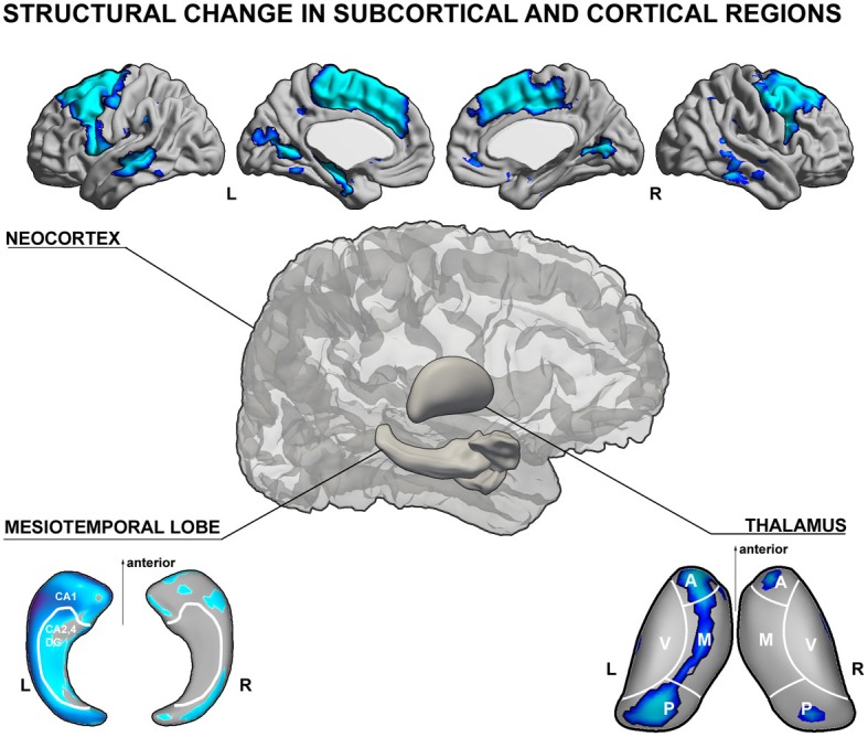
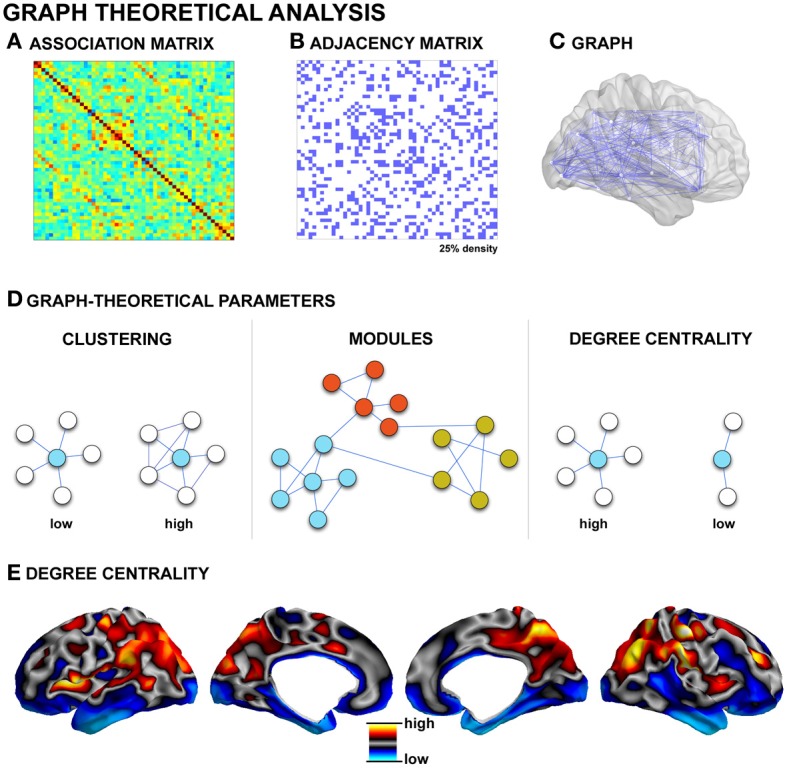
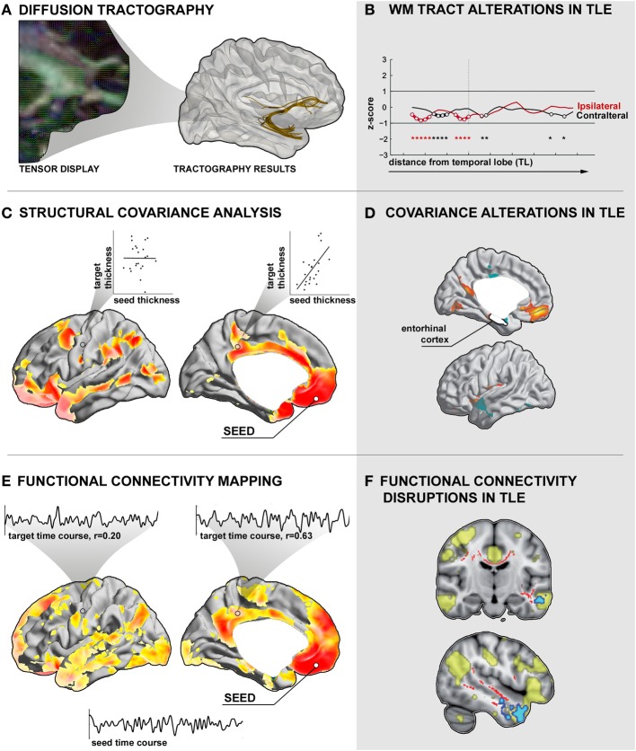
Early imaging studies in temporal lobe epilepsy (TLE) focused on the search for mesial temporal sclerosis, as its surgical removal results in clinically meaningful improvement in about 70% of patients. Nevertheless, a considerable subgroup of patients continues to suffer from post-operative seizures. Although the reasons for surgical failure are not fully understood, electrophysiological and imaging data suggest that anomalies extending beyond the temporal lobe may have negative impact on outcome. This hypothesis has revived the concept of human epilepsy as a disorder of distributed brain networks. Recent methodological advances in non-invasive neuroimaging have led to quantify structural and functional networks in vivo. While structural networks can be inferred from diffusion MRI tractography and inter-regional covariance patterns of structural measures such as cortical thickness, functional connectivity is generally computed based on statistical dependencies of neurophysiological time-series, measured through functional MRI or electroencephalographic techniques. This review considers the application of advanced analytical methods in structural and functional connectivity analyses in TLE. We will specifically highlight findings from graph-theoretical analysis that allow assessing the topological organization of brain networks. These studies have provided compelling evidence that TLE is a system disorder with profound alterations in local and distributed networks. In addition, there is emerging evidence for the utility of network properties as clinical diagnostic markers. Nowadays, a network perspective is considered to be essential to the understanding of the development, progression, and management of epilepsy.
早期针对颞叶癫痫(TLE)的影像学研究主要聚焦于寻找内侧颞叶硬化,因为手术切除该区域能使约70%的患者在临床上获得显著改善。然而,仍有相当一部分患者术后继续遭受癫痫发作。尽管手术失败的原因尚未完全明确,但电生理和影像学数据表明,超出颞叶范围的异常可能会对手术结果产生负面影响。这一假说重新唤起了人们对于人类癫痫是一种分布式脑网络疾病的认识。非侵入性神经影像学的最新方法进展已能够在活体中对结构和功能网络进行量化。结构网络可通过扩散磁共振成像纤维束成像以及诸如皮质厚度等结构测量的区域间协方差模式来推断,而功能连接性通常是基于神经生理时间序列的统计依赖性来计算的,这些时间序列通过功能磁共振成像或脑电图技术进行测量。本综述探讨了先进分析方法在TLE结构和功能连接性分析中的应用。我们将特别强调来自图论分析的结果,这些结果有助于评估脑网络的拓扑组织。这些研究提供了令人信服的证据,表明TLE是一种系统疾病,局部和分布式网络存在深刻改变。此外,越来越多的证据表明网络属性作为临床诊断标志物具有实用性。如今,网络视角被认为对于理解癫痫的发生、发展和管理至关重要。