Laboratório de Pesquisa em Leishmaniose, Instituto Oswaldo Cruz, FIOCRUZ, Rio de Janeiro, Brasil ; Laboratório de Biologia Molecular e Doenças Endêmicas, Instituto Oswaldo Cruz, FIOCRUZ, Rio de Janeiro, Brasil.
PLoS Negl Trop Dis. 2013 Oct 17;7(10):e2481. doi: 10.1371/journal.pntd.0002481. eCollection 2013.
Iron is an essential element for the survival of microorganisms in vitro and in vivo, acting as a cofactor of several enzymes and playing a critical role in host-parasite relationships. Leishmania (Viannia) braziliensis is a parasite that is widespread in the new world and considered the major etiological agent of American tegumentary leishmaniasis. Although iron depletion leads to promastigote and amastigote growth inhibition, little is known about the role of iron in the biology of Leishmania. Furthermore, there are no reports regarding the importance of iron for L. (V.) braziliensis.
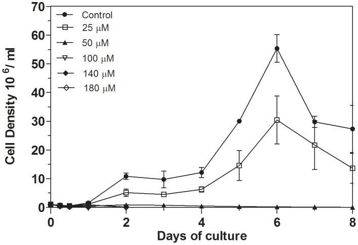
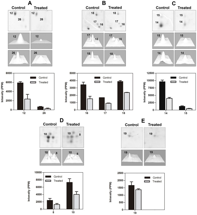
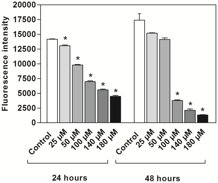
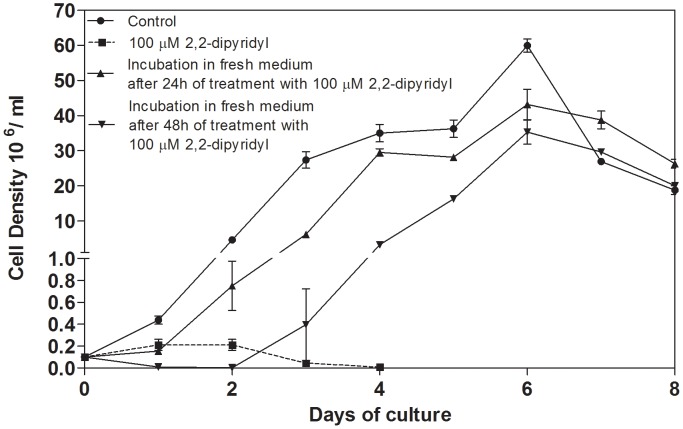
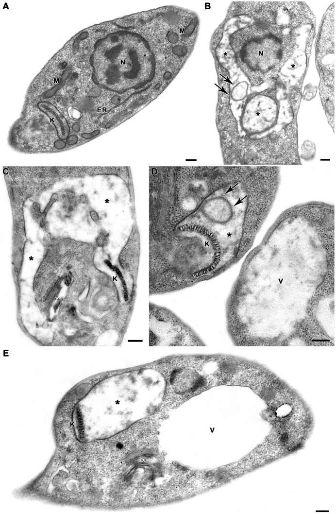
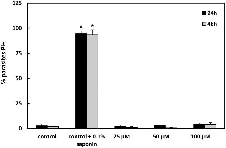
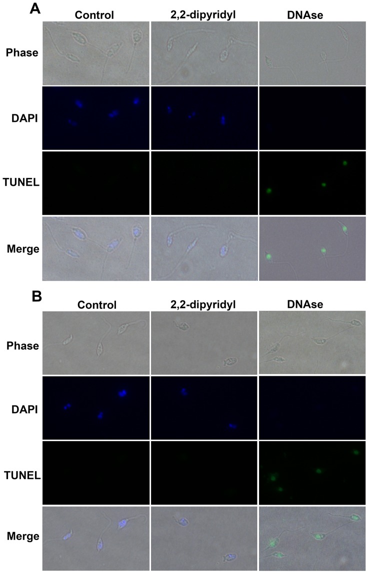
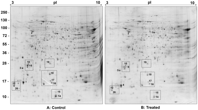
METHODOLOGY/PRINCIPAL FINDINGS: In this study, the effect of iron on the growth, ultrastructure and protein expression of L. (V.) braziliensis was analyzed by the use of the chelator 2,2-dipyridyl. Treatment with 2,2-dipyridyl affected parasites' growth in a dose- and time-dependent manner. Multiplication of the parasites was recovered after reinoculation in fresh culture medium. Ultrastructural analysis of treated promastigotes revealed marked mitochondrial swelling with loss of cristae and matrix and the presence of concentric membranar structures inside the organelle. Iron depletion also induced Golgi disruption and intense cytoplasmic vacuolization. Fluorescence-activated cell sorting analysis of tetramethylrhodamine ester-stained parasites showed that 2,2-dipyridyl collapsed the mitochondrial membrane potential. The incubation of parasites with propidium iodide demonstrated that disruption of mitochondrial membrane potential was not associated with plasma membrane permeabilization. TUNEL assays indicated no DNA fragmentation in chelator-treated promastigotes. In addition, two-dimensional electrophoresis showed that treatment with the iron chelator induced up- or down-regulation of proteins involved in metabolism of nucleic acids and coordination of post-translational modifications, without altering their mRNA levels.
Iron chelation leads to a multifactorial response that results in cellular collapse, starting with the interruption of cell proliferation and culminating in marked mitochondrial impairment in some parasites and their subsequent cell death, whereas others may survive and resume proliferating.
铁是微生物在体外和体内生存的必需元素,作为几种酶的辅助因子,在宿主-寄生虫关系中起着关键作用。巴西利什曼原虫(Viannia)是一种广泛存在于新世界的寄生虫,被认为是美洲皮肤利什曼病的主要病原体。尽管铁耗竭会导致前鞭毛体和无鞭毛体的生长抑制,但人们对铁在利什曼原虫生物学中的作用知之甚少。此外,尚无关于铁对 L.(V.)braziliensis 重要性的报道。
方法/主要发现:在这项研究中,使用螯合剂 2,2-二吡啶来分析铁对 L.(V.)braziliensis 的生长、超微结构和蛋白质表达的影响。2,2-二吡啶的处理以剂量和时间依赖的方式影响寄生虫的生长。在新鲜培养基中重新接种后,寄生虫的繁殖得到恢复。用处理过的前鞭毛体进行的超微结构分析显示,线粒体明显肿胀,嵴和基质丢失,细胞器内存在同心膜状结构。铁耗竭还诱导高尔基体破坏和细胞质强烈空泡化。用四甲基罗丹明乙酯染色的寄生虫荧光激活细胞分选分析表明,2,2-二吡啶使线粒体膜电位崩溃。用碘化丙啶孵育寄生虫表明,线粒体膜电位的破坏与质膜通透性的破坏无关。TUNEL 测定表明螯合剂处理的前鞭毛体中没有 DNA 片段化。此外,二维电泳显示,用铁螯合剂处理会引起参与核酸代谢和翻译后修饰协调的蛋白质的上调或下调,而不会改变其 mRNA 水平。
铁螯合作用导致多因素反应,导致细胞崩溃,首先是细胞增殖中断,最终导致一些寄生虫明显的线粒体损伤及其随后的细胞死亡,而其他寄生虫可能存活并恢复增殖。