Division of Virology, National Institute for Biological Standards and Control, Medicines and Healthcare products Regulatory Agency, Blanche Lane, South Mimms, Potters Bar, Hertfordshire EN6 3QG, UK.
Internal Medicine Research Unit, Pfizer Research and Development, Sandwich, Kent CT13 9NJ, UK.
Virus Res. 2014 Jan 22;179(100):93-101. doi: 10.1016/j.virusres.2013.11.006. Epub 2013 Nov 15.
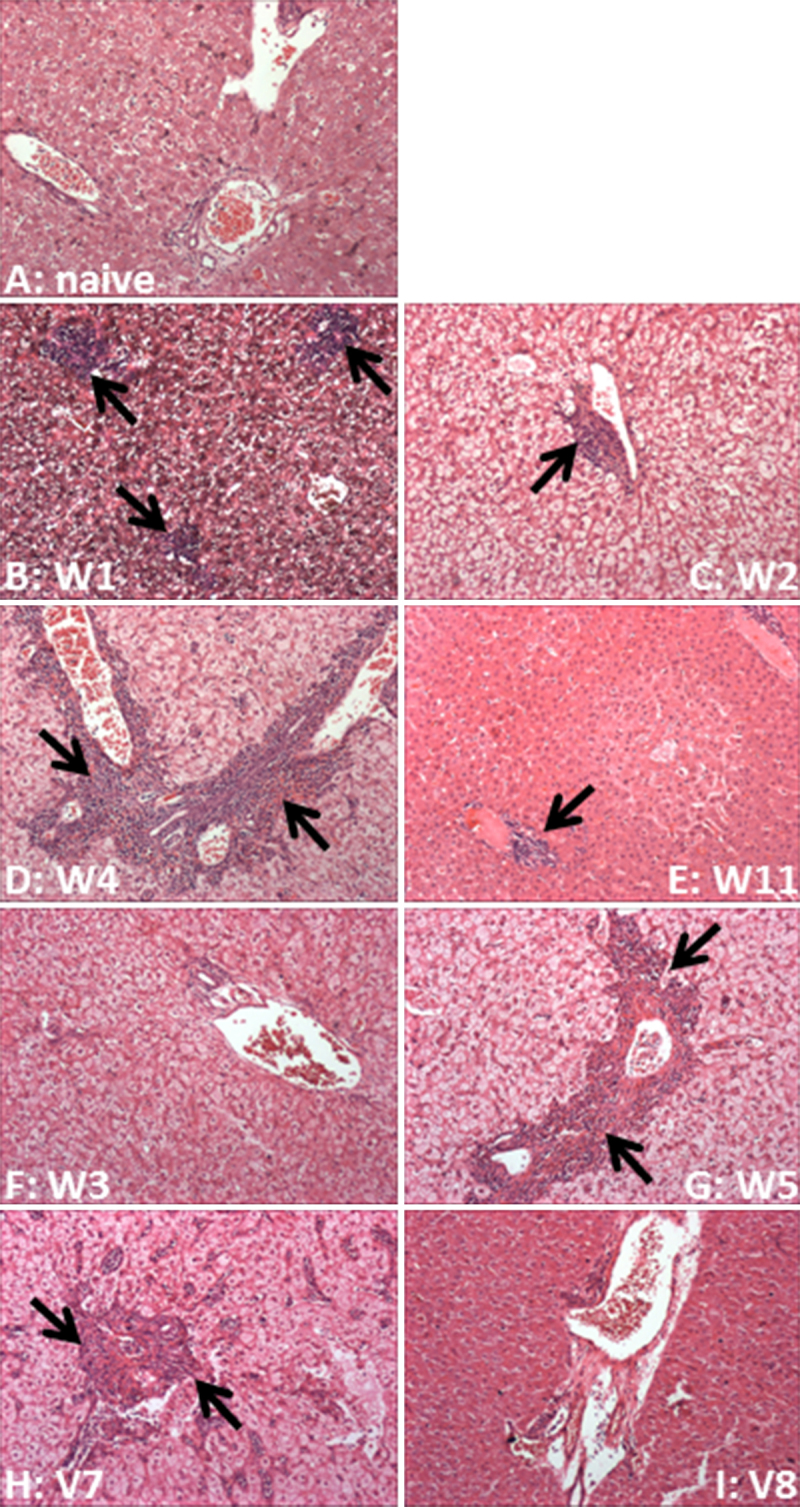
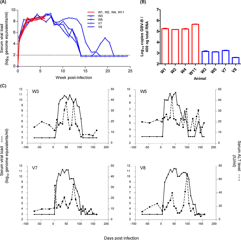
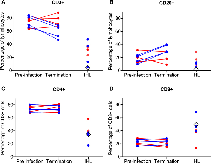
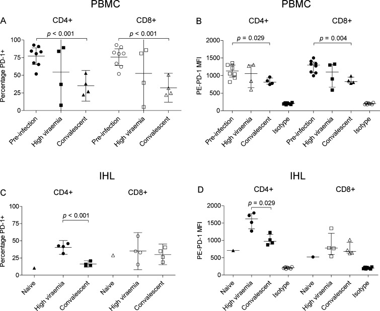
Flaviviruses related to hepatitis C virus (HCV) in suitable animal models may provide further insight into the role that cellular immunity contributes to spontaneous clearance of HCV. We characterised changes in lymphocyte populations in tamarins with an acute GBV-B infection, a hepatitis virus of the flaviviridae. Major immune cell populations were monitored in peripheral and intra-hepatic lymphocytes at high viraemia or following a period when peripheral virus was no longer detected. Limited changes in major lymphocyte populations were apparent during high viraemia; however, the proportions of CD3(+) lymphocytes decreased and CD20(+) lymphocytes increased once peripheral viraemia became undetectable. Intrahepatic lymphocyte populations increased at both time points post-infection. Distinct expression patterns of PD-1, a marker of T-cell activation, were observed on peripheral and hepatic lymphocytes; notably there was elevated PD-1 expression on hepatic CD4(+) T-cells during high viraemia, suggesting an activated phenotype, which decreased following clearance of peripheral viraemia. At times when peripheral vRNA was not detected, suggesting viral clearance, we were able to readily detect GBV-B RNA in the liver, indicative of long-term virus replication. This study is the first description of changes in lymphocyte populations during GBV-B infection of tamarins and provides a foundation for more detailed investigations of the responses that contribute to the control of GBV-B infection.
在合适的动物模型中,与丙型肝炎病毒 (HCV) 相关的黄病毒可能会进一步深入了解细胞免疫在 HCV 自发性清除中所起的作用。我们对感染急性 GBV-B(黄病毒科的一种肝炎病毒)的绢毛猴的淋巴细胞群变化进行了特征描述。在高病毒血症或外周病毒检测不到后,监测外周和肝内淋巴细胞中的主要免疫细胞群。在高病毒血症期间,主要淋巴细胞群的变化有限;然而,一旦外周病毒血症检测不到,CD3(+) 淋巴细胞的比例就会下降,CD20(+) 淋巴细胞的比例就会增加。感染后两个时间点肝内淋巴细胞群均增加。在外周和肝淋巴细胞上观察到 PD-1(T 细胞活化的标志物)的表达模式明显不同;值得注意的是,在高病毒血症期间,肝 CD4(+) T 细胞上的 PD-1 表达升高,表明存在激活表型,随着外周病毒血症清除,其表达降低。在外周 vRNA 检测不到,提示病毒清除时,我们能够在肝脏中轻易检测到 GBV-B RNA,表明长期病毒复制。本研究首次描述了 GBV-B 感染绢毛猴期间淋巴细胞群的变化,并为更详细地研究控制 GBV-B 感染的反应提供了基础。