Respiratory Division, University Hospital Leuven, Department of Clinical and Experimental Medicine, KU Leuven, Leuven, Belgium.
Respir Res. 2013 Nov 19;14(1):131. doi: 10.1186/1465-9921-14-131.
Spirometric parameters are the mainstay for diagnosis of COPD, but cannot distinguish airway obstruction from emphysema. We aimed to develop a computer model that quantifies airway collapse on forced expiratory flow-volume loops. We then explored and validated the relationship of airway collapse with computed tomography (CT) diagnosed emphysema in two large independent cohorts.
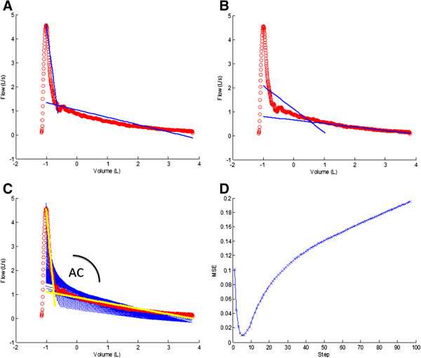
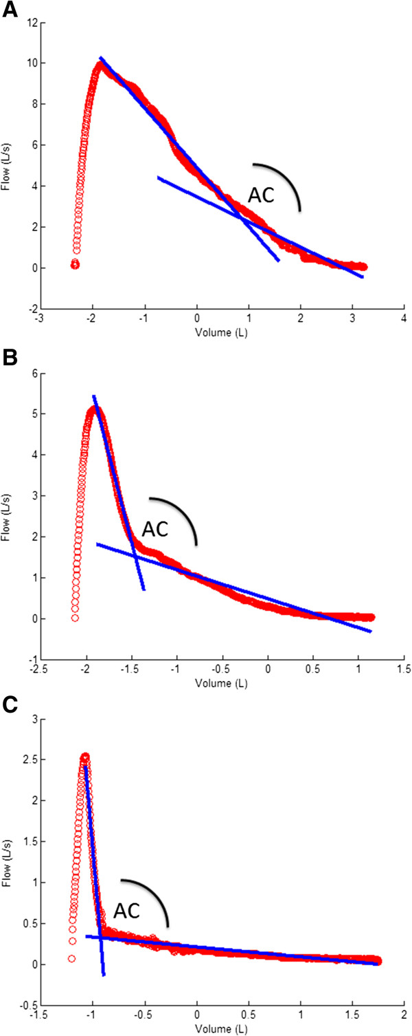
A computer model was developed in 513 Caucasian individuals with ≥15 pack-years who performed spirometry, diffusion capacity and CT scans to quantify emphysema presence. The model computed the two best fitting regression lines on the expiratory phase of the flow-volume loop and calculated the angle between them. The collapse was expressed as an Angle of collapse (AC) which was then correlated with the presence of emphysema. Findings were validated in an independent group of 340 individuals.
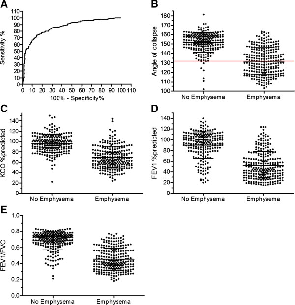
AC in emphysema subjects (N = 251) was significantly lower (131° ± 14°) compared to AC in subjects without emphysema (N = 223), (152° ± 10°) (p < 0.0001). Multivariate regression analysis revealed AC as best indicator of visually scored emphysema (R2 = 0.505, p < 0.0001) with little significant contribution of KCO, %predicted and FEV1, %predicted to the total model (total R2 = 0.626, p < 0.0001). Similar associations were obtained when using CT-automated density scores for emphysema assessment. Receiver operating characteristic (ROC) curves pointed to 131° as the best cut-off for emphysema (95.5% positive predictive value, 97% specificity and 51% sensitivity). Validation in a second group confirmed the significant difference in mean AC between emphysema and non-emphysema subjects. When applying the 131° cut-off, a positive predictive value of 95.6%, a specificity of 96% and a sensitivity of 59% were demonstrated.
Airway collapse on forced expiration quantified by a computer model correlates with emphysema. An AC below 131° can be considered as a specific cut-off for predicting the presence of emphysema in heavy smokers.
肺量测定参数是诊断 COPD 的主要依据,但无法区分气道阻塞与肺气肿。我们旨在开发一种能够量化用力呼气流量容积环中气道塌陷的计算机模型。然后,我们在两个大型独立队列中探索并验证了气道塌陷与 CT 诊断肺气肿之间的关系。
在 513 名至少吸 15 包年烟的白种人中建立了一个计算机模型,他们进行了肺量测定、弥散能力和 CT 扫描以量化肺气肿的存在。该模型在流量容积环的呼气阶段计算出两条最佳拟合回归线,并计算它们之间的夹角。塌陷表示为塌陷角(AC),然后与肺气肿的存在相关联。在 340 名独立个体中验证了该模型。
肺气肿组(N=251)的 AC 明显低于无肺气肿组(N=223)(131°±14°)(p<0.0001)。多元回归分析显示,AC 是视觉评分肺气肿的最佳指标(R2=0.505,p<0.0001),KCO、%预计值和 FEV1、%预计值对总模型的贡献很小(总 R2=0.626,p<0.0001)。使用 CT 自动密度评分评估肺气肿时也得到了类似的关联。受试者工作特征(ROC)曲线显示 131°为最佳肺气肿截断值(95.5%阳性预测值、97%特异性和 51%敏感性)。在第二组中的验证证实了肺气肿和非肺气肿组之间 AC 的平均值存在显著差异。当应用 131°截断值时,阳性预测值为 95.6%,特异性为 96%,敏感性为 59%。
通过计算机模型量化的用力呼气时气道塌陷与肺气肿相关。AC 低于 131°可作为预测重度吸烟者肺气肿存在的特定截断值。