Puri Claudia, Renna Maurizio, Bento Carla Figueira, Moreau Kevin, Rubinsztein David C
Department of Medical Genetics; Cambridge Institute for Medical Research; University of Cambridge; Cambridge UK.
Autophagy. 2014 Jan;10(1):182-4. doi: 10.4161/auto.27174. Epub 2013 Nov 19.
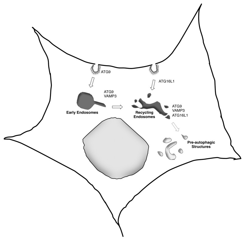
Autophagosomes are formed by double-membraned structures, which engulf portions of cytoplasm. Autophagosomes ultimately fuse with lysosomes, where their contents are degraded. The origin of the autophagosome membrane may involve different sources, such as mitochondria, Golgi, endoplasmic reticulum, plasma membrane, and recycling endosomes. We recently observed that ATG9 localizes on the plasma membrane in clathrin-coated structures and is internalized following a classical endocytic pathway through early and then recycling endosomes. By contrast, ATG16L1 is also internalized by clathrin-mediated endocytosis but via different clathrin-coated pits, and appears to follow a different route to the recycling endosomes. The R-SNARE VAMP3 mediates the coalescence of the 2 different pools of vesicles (containing ATG16L1 or ATG9) in recycling endosomes. The heterotypic fusion between ATG16L1- and ATG9-containing vesicles strongly correlates with subsequent autophagosome formation. Thus, ATG9 and ATG16L1 both traffic from the plasma membrane to autophagic precursor structures and provide 2 routes from the plasma membrane to autophagosomes.
自噬体由双膜结构形成,其包裹部分细胞质。自噬体最终与溶酶体融合,在溶酶体中其内容物被降解。自噬体膜的起源可能涉及不同来源,如线粒体、高尔基体、内质网、质膜和再循环内体。我们最近观察到,ATG9定位于网格蛋白包被结构中的质膜上,并通过经典的内吞途径,先经早期再循环内体然后经再循环内体被内化。相比之下,ATG16L1也通过网格蛋白介导的内吞作用被内化,但通过不同的网格蛋白包被小窝,并且似乎遵循不同的途径到达再循环内体。R-SNARE蛋白VAMP3介导再循环内体中两种不同囊泡池(含有ATG16L1或ATG9)的融合。含有ATG16L1和ATG9的囊泡之间的异型融合与随后的自噬体形成密切相关。因此,ATG9和ATG16L1都从质膜运输到自噬前体结构,并提供了从质膜到自噬体的两条途径。