Department of Psychiatry and Behavioral Sciences, University of Southern California, Los Angeles, California, United States of America.
PLoS One. 2013 Nov 21;8(11):e80058. doi: 10.1371/journal.pone.0080058. eCollection 2013.
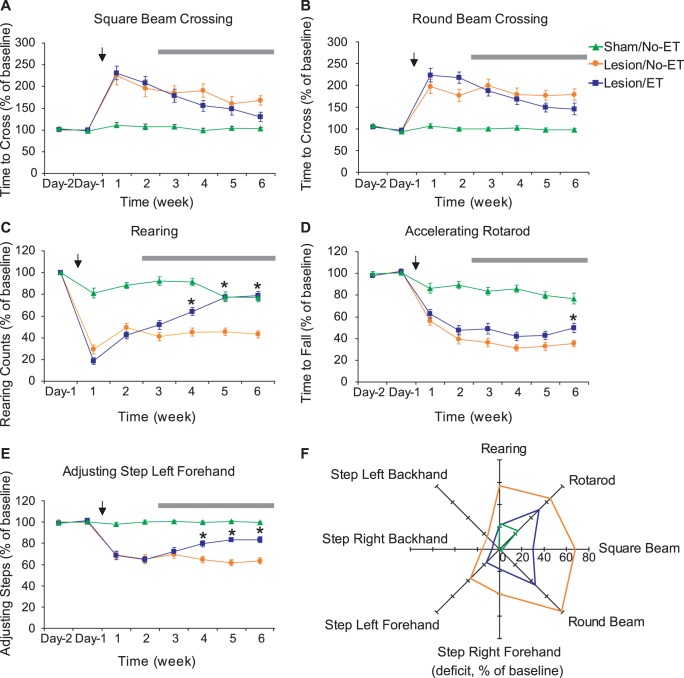
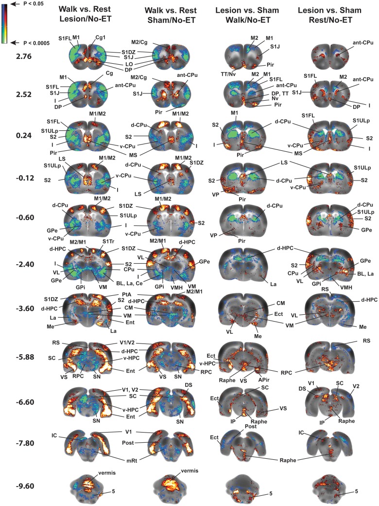
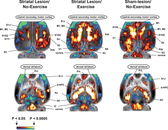
Exercise training is widely used for neurorehabilitation of Parkinson's disease (PD). However, little is known about the functional reorganization of the injured brain after long-term aerobic exercise. We examined the effects of 4 weeks of forced running wheel exercise in a rat model of dopaminergic deafferentation (bilateral, dorsal striatal 6-hydroxydopamine lesions). One week after training, cerebral perfusion was mapped during treadmill walking or at rest using [(14)C]-iodoantipyrine autoradiography. Regional cerebral blood flow-related tissue radioactivity (rCBF) was analyzed in three-dimensionally reconstructed brains by statistical parametric mapping. In non-exercised rats, lesions resulted in persistent motor deficits. Compared to sham-lesioned rats, lesioned rats showed altered functional brain activation during walking, including: 1. hypoactivation of the striatum and motor cortex; 2. hyperactivation of non-lesioned areas in the basal ganglia-thalamocortical circuit; 3. functional recruitment of the red nucleus, superior colliculus and somatosensory cortex; 4. hyperactivation of the ventrolateral thalamus, cerebellar vermis and deep nuclei, suggesting recruitment of the cerebellar-thalamocortical circuit; 5. hyperactivation of limbic areas (amygdala, hippocampus, ventral striatum, septum, raphe, insula). These findings show remarkable similarities to imaging findings reported in PD patients. Exercise progressively improved motor deficits in lesioned rats, while increasing activation in dorsal striatum and rostral secondary motor cortex, attenuating a hyperemia of the zona incerta and eliciting a functional reorganization of regions participating in the cerebellar-thalamocortical circuit. Both lesions and exercise increased activation in mesolimbic areas (amygdala, hippocampus, ventral striatum, laterodorsal tegmental n., ventral pallidum), as well as in related paralimbic regions (septum, raphe, insula). Exercise, but not lesioning, resulted in decreases in rCBF in the medial prefrontal cortex (cingulate, prelimbic, infralimbic). Our results in this PD rat model uniquely highlight the breadth of functional reorganizations in motor and limbic circuits following lesion and long-term, aerobic exercise, and provide a framework for understanding the neural substrates underlying exercise-based neurorehabilitation.
运动训练被广泛应用于帕金森病(PD)的神经康复。然而,对于长期有氧运动后受损大脑的功能重组,我们知之甚少。我们在多巴胺能去传入大鼠模型(双侧,背侧纹状体 6-羟多巴胺损伤)中检查了 4 周强制跑步轮运动的效果。在训练后 1 周,使用 [(14)C]-碘安替比林放射自显影术在跑步机行走或休息期间绘制脑灌注图。通过统计参数映射,在三维重建大脑中分析与区域脑血流相关的组织放射性(rCBF)。在未运动的大鼠中,损伤导致持续的运动缺陷。与假损伤大鼠相比,损伤大鼠在行走过程中表现出改变的功能性大脑激活,包括:1. 纹状体和运动皮层的低激活;2. 基底节-丘脑皮质回路中非损伤区域的高激活;3. 红核、上丘和躯体感觉皮层的功能募集;4. 腹外侧丘脑、小脑蚓部和深部核团的高激活,提示小脑-丘脑皮质回路的募集;5. 边缘区域(杏仁核、海马体、腹侧纹状体、隔区、中缝核、脑岛)的高激活。这些发现与 PD 患者的影像学发现非常相似。运动逐渐改善了损伤大鼠的运动缺陷,同时增加了背侧纹状体和初级运动皮层的激活,减轻了未定带的充血,并引起了参与小脑-丘脑皮质回路的区域的功能重组。损伤和运动都增加了中边缘区域(杏仁核、海马体、腹侧纹状体、外侧背侧被盖核、腹侧苍白球)以及相关的边缘区域(隔区、中缝核、脑岛)的激活。运动,而不是损伤,导致内侧前额叶皮质(扣带回、前扣带回、下边缘回)的 rCBF 降低。我们在这个 PD 大鼠模型中的结果独特地突出了运动和边缘回路在损伤和长期有氧运动后的广泛功能重组,并为理解基于运动的神经康复的神经基础提供了框架。