Academic Unit of Ophthalmology, Centre for Translational Inflammation Research, University of Birmingham, Birmingham, United Kingdom.
Wolfson Computer Laboratory, Queen Elizabeth Hospital, Birmingham, United Kingdom.
Ophthalmology. 2014 Feb;121(2):492-7. doi: 10.1016/j.ophtha.2013.09.033. Epub 2013 Dec 4.
Quantifying the extent of conjunctival fibrosis for documentation of progression in conjunctival scarring disease is a clinical challenge. Measurement of forniceal foreshortening facilitates monitoring of these disorders. This study aims (1) to define the limits of the normal human conjunctival fornices and how these alter with age and (2) to provide normative data for upper and lower fornix depths (FDs) and fornix intercanthal distance (FICD) within a healthy South Asian, racially distinct population.
Epidemiologic, cross-sectional study.
A total of 240 subjects with national origins from South Asia, with no known ocular history and normal adnexal and conjunctival examination, aged 20 to 80 years.
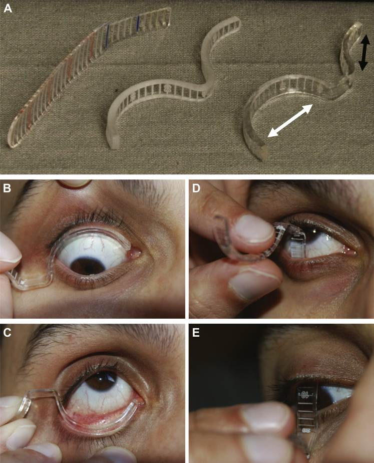
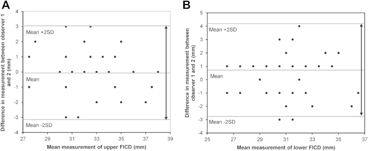
An FICD modification of a custom-designed fornix depth measurer (FDM) was validated and used for measurement of both lower and upper FDs together with FICDs in 480 healthy eyes with no ocular comorbidities. Data were analyzed using repeated-measures analysis of variance and presented as means with 95% confidence intervals (CIs).
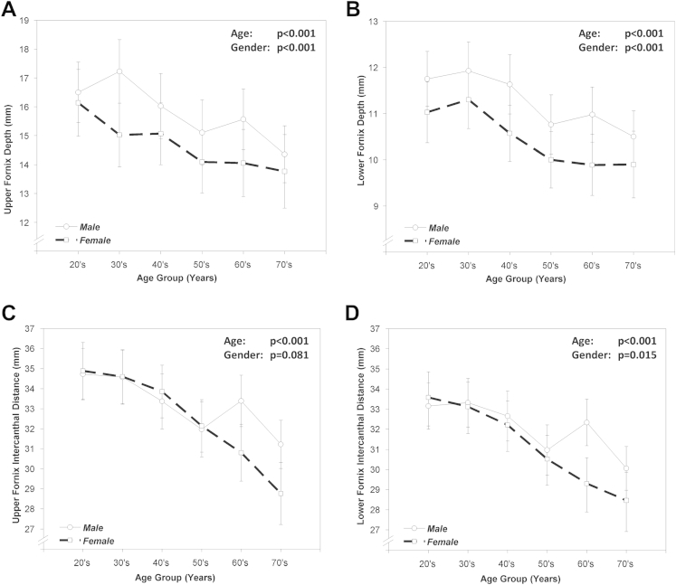
Mean lower and upper FDs and FICD for the entire cohort, stratified according to age decade and sex.
For this South Asian population, the overall upper and lower FDs were 15.3 mm (95% CI, 14.9-15.6) and 10.9 mm (95% CI, 10.7-11.1), respectively, with FICD defined as 32.9 mm (95% CI, 32.5-33.4) (upper) and 31.7 mm (95% CI, 31.3-32.1) (lower). With increasing age, a progressive reduction of all measured parameters (P < 0.001) was noted, with female subjects having significantly shallower fornices (upper FD, P < 0.001; lower FD, P < 0.001; upper FICD, P = 0.081; and lower FICD, P = 0.015).
This is the first study to define the limits of normal upper FD and FICDs in any population group. Our study demonstrates sex variations and progressive conjunctival shrinkage with age. Although it provides important, objective data for normal forniceal anatomy, further study is recommended in other populations to confirm the generalizability of these data or to enable normal comparative datasets for the assessment of conjunctival scarring disorders among all anthropological groups.
定量评估结膜纤维化的程度,以记录结膜瘢痕疾病的进展,这在临床上是一个挑战。穹窿缩短的测量有助于监测这些疾病。本研究旨在:(1)确定正常人类结膜穹窿的范围以及这些范围如何随年龄变化而变化;(2)为健康的南亚人群中的上穹窿和下穹窿深度(FD)以及穹窿内眦距离(FICD)提供正常参考值。
流行病学、横断面研究。
共 240 名受试者,来自南亚各国,无眼部疾病史,附属器和结膜检查正常,年龄 20 至 80 岁。
使用改良的定制穹窿深度测量仪(FDM)对 FICD 进行了验证,并用于测量 480 只无眼部合并症的健康眼睛的下穹窿和上穹窿 FD 以及 FICD。采用重复测量方差分析对数据进行分析,并以均数和 95%置信区间(CI)表示。
整个队列的平均下穹窿和上穹窿 FD 以及 FICD,按年龄十年和性别分层。
对于这个南亚人群,总上穹窿和下穹窿 FD 分别为 15.3 毫米(95%CI,14.9-15.6)和 10.9 毫米(95%CI,10.7-11.1),FICD 定义为 32.9 毫米(95%CI,32.5-33.4)(上)和 31.7 毫米(95%CI,31.3-32.1)(下)。随着年龄的增长,所有测量参数均呈渐进性下降(P<0.001),女性受试者的穹窿明显较浅(上穹窿 FD,P<0.001;下穹窿 FD,P<0.001;上 FICD,P=0.081;下 FICD,P=0.015)。
这是第一项在任何人群中定义正常上穹窿 FD 和 FICD 范围的研究。本研究表明存在性别差异,并随年龄增长出现进行性结膜收缩。尽管为正常穹窿解剖结构提供了重要的客观数据,但建议在其他人群中进一步研究,以确认这些数据的普遍性,或为所有人类学群体评估结膜瘢痕疾病提供正常的对照数据集。