Athinoula A. Martinos Center for Biomedical Imaging, Massachusetts General Hospital, 149 Thirteenth Street, Suite 2301, Charlestown, MA 02129, USA.
Division of Epilepsy and Clinical Neurophysiology, Boston Children's Hospital, 300 Longwood Avenue, Boston, MA 02115, USA.
Epilepsy Res. 2014 Feb;108(2):280-8. doi: 10.1016/j.eplepsyres.2013.11.006. Epub 2013 Nov 18.
To investigate the correlation between spike propagation represented by spatiotemporal source analysis of magnetoencephalographic (MEG) spikes and surgical outcome in patients with temporal lobe epilepsy.
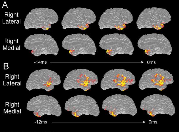
Thirty-seven patients were divided into mesial (n=27) and non-mesial (n=10) groups based on the presurgical evaluation. In each patient, ten ipsilateral spikes were averaged, and spatiotemporal source maps of the averaged spike were obtained by using minimum norm estimate. Regions of interest (ROIs) were created including temporoparietal, inferior frontal, mesial temporal, anterior and posterior part of the lateral temporal cortex. We extracted activation values from the source maps and the threshold was set at half of the maximum activation at the peak latency. The leading and propagated areas of the spike were defined as those ROIs with activation reaching the threshold at the earliest and at the peak latencies, respectively. Surgical outcome was assessed based on Engel's classification. Binary variables were created from leading areas (restricted to the anterior and mesial temporal ROIs or not) and from propagation areas (involving the temporoparietal ROI or not), and for surgical outcome (Class I or not). Fisher's exact test was used for significance testing.
In total and mesial group, restricted anterior/mesial temporal leading areas were correlated with Class I (p<0.05). Temporoparietal propagation was correlated with Class II-IV (p<0.05). For the non-mesial group, no significant relation was found.
Spike propagation patterns represented by spatiotemporal source analysis of MEG spikes may provide useful information for prognostic implication in presurgical evaluation of epilepsy.
研究经时源分析的脑磁图(MEG)棘波时空传播与颞叶癫痫患者手术结果的相关性。
根据术前评估,将 37 例患者分为内侧(n=27)和非内侧(n=10)组。在每位患者中,平均 10 个同侧棘波,并用最小范数估计获得平均棘波的时空源图。创建感兴趣区域(ROI),包括颞顶叶、下额叶、内侧颞叶、外侧颞叶的前、后部分。我们从源图中提取激活值,并将阈值设置为最大激活值在峰潜伏期的一半。棘波的起始和传播区域定义为在最早和峰潜伏期达到阈值的 ROI。手术结果根据恩格尔分类进行评估。创建来自引导区域(仅限于前内侧颞叶 ROI 或不)和传播区域(涉及颞顶叶 ROI 或不)的二项变量,并与手术结果(I 级或非 I 级)进行比较。使用 Fisher 确切检验进行显著性检验。
在所有患者和内侧组中,限制在前/内侧颞叶的引导区域与 I 级(p<0.05)相关。颞顶叶的传播与 II-IV 级(p<0.05)相关。对于非内侧组,未发现显著相关性。
MEG 棘波的时空源分析表示的棘波传播模式可能为癫痫术前评估的预后提供有用信息。