Schormann N, Banerjee S, Ricciardi R, Chattopadhyay D
Department of Medicine, University of Alabama at Birmingham, Birmingham, AL 35294, USA.
Acta Crystallogr Sect F Struct Biol Cryst Commun. 2013 Dec;69(Pt 12):1328-34. doi: 10.1107/S1744309113030613. Epub 2013 Nov 28.
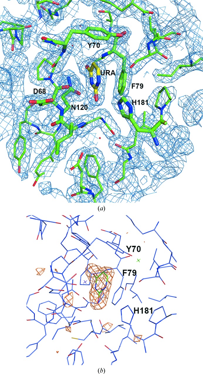
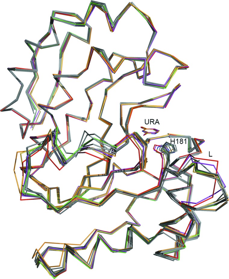
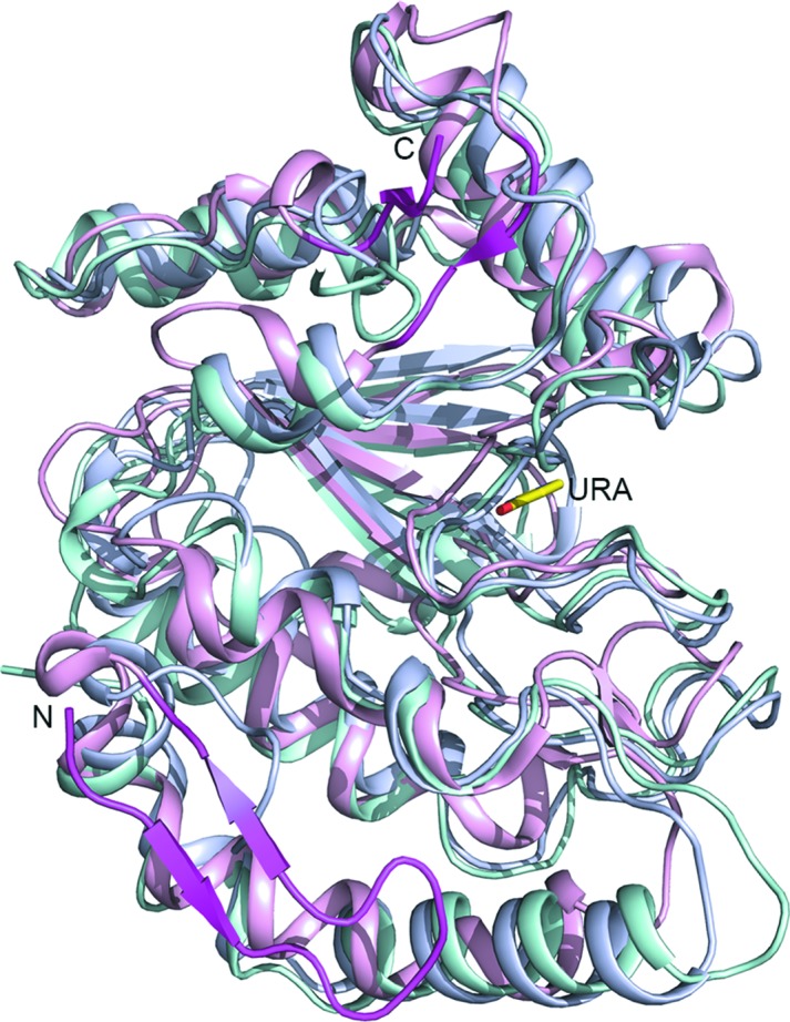
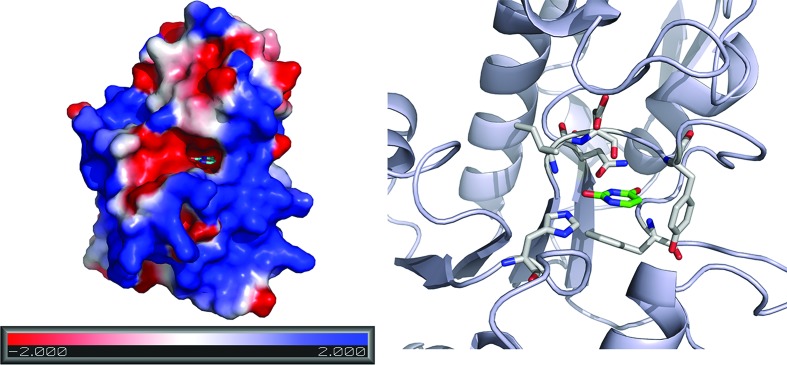
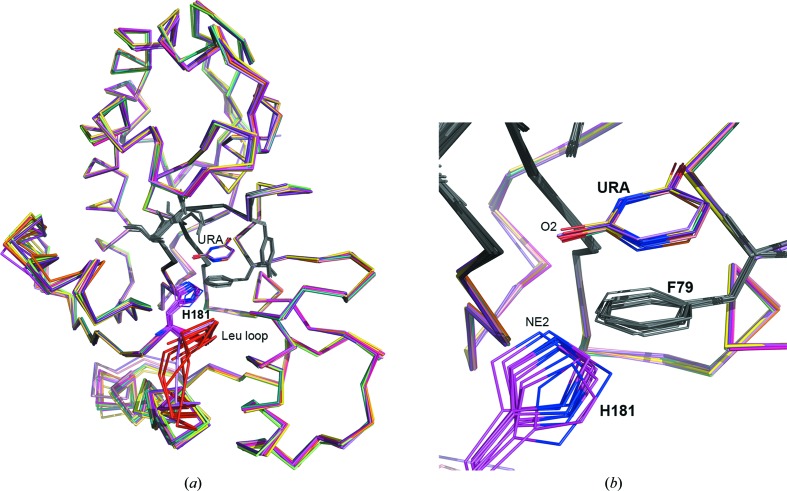
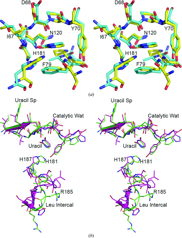
Poxvirus uracil DNA glycosylases are the most diverse members of the family I uracil DNA glycosylases (UNGs). The crystal structure of the uracil complex of Vaccinia virus uracil DNA glycosylase (D4) was determined at 2.03 Å resolution. One uracil molecule was located in the active-site pocket in each of the 12 noncrystallographic symmetry-related D4 subunits. Although the UNGs of the poxviruses (including D4) feature significant differences in the characteristic motifs designated for uracil recognition and in the base-excision mechanism, the architecture of the active-site pocket in D4 is very similar to that in UNGs of other organisms. Overall, the interactions of the bound uracil with the active-site residues are also similar to the interactions previously observed in the structures of human and Escherichia coli UNG.
痘病毒尿嘧啶DNA糖基化酶是I型尿嘧啶DNA糖基化酶(UNG)家族中最多样化的成员。痘苗病毒尿嘧啶DNA糖基化酶(D4)的尿嘧啶复合物的晶体结构在2.03 Å分辨率下得以确定。在12个非晶体学对称相关的D4亚基的每一个中,一个尿嘧啶分子位于活性位点口袋中。尽管痘病毒的UNG(包括D4)在用于尿嘧啶识别的特征基序以及碱基切除机制方面存在显著差异,但D4中活性位点口袋的结构与其他生物体UNG中的结构非常相似。总体而言,结合的尿嘧啶与活性位点残基的相互作用也与先前在人类和大肠杆菌UNG结构中观察到的相互作用相似。